Copyright
©2008 The WJG Press and Baishideng.
World J Gastroenterol. Mar 14, 2008; 14(10): 1575-1581
Published online Mar 14, 2008. doi: 10.3748/wjg.14.1575
Published online Mar 14, 2008. doi: 10.3748/wjg.14.1575
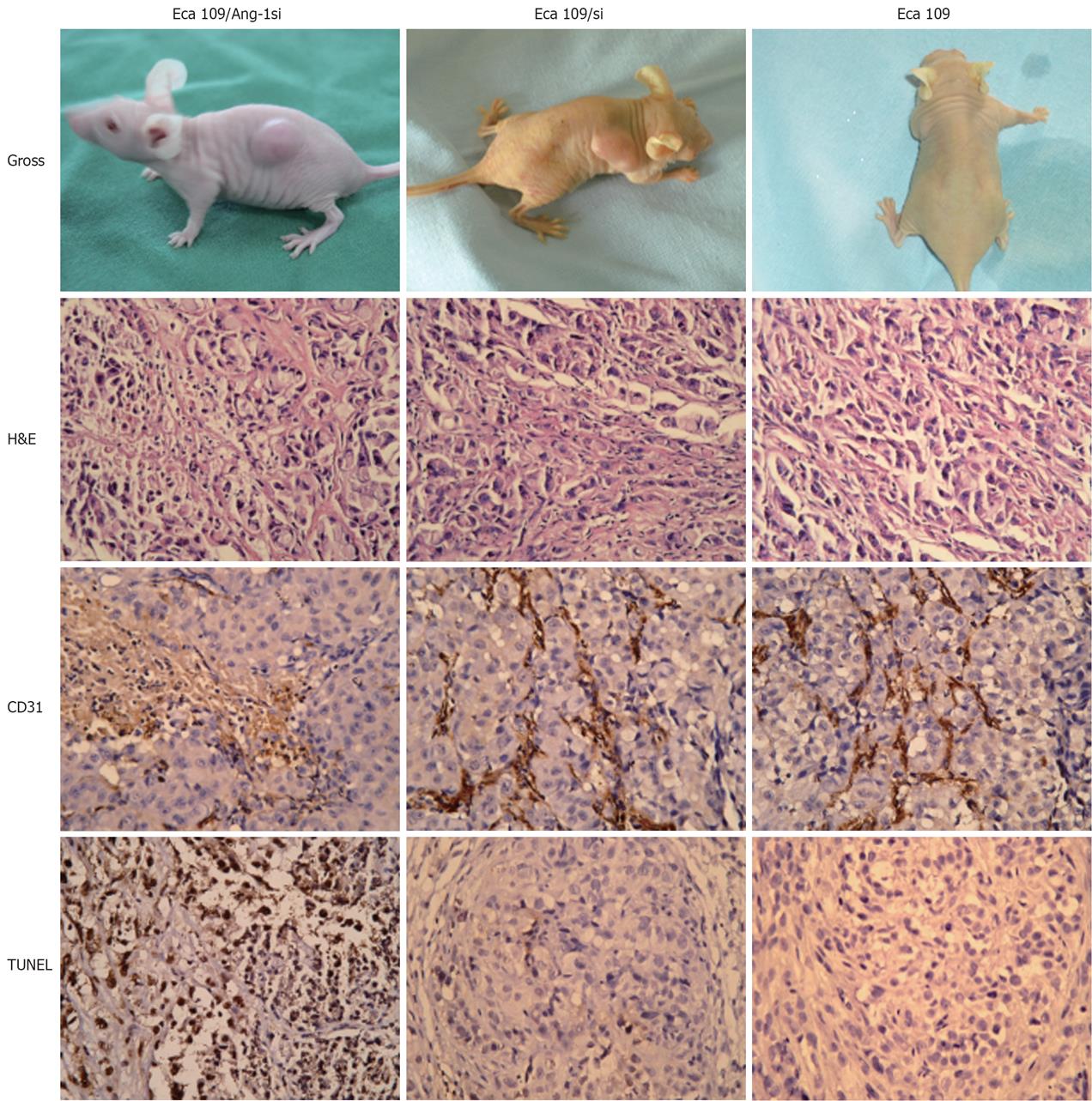
Figure 3 Effect of Ang-1si on esophageal cancer growth in vivo.
Grossly, the Eca109/Ang-1si tumors were significantly smaller than those in both control groups; Under microscopy, massive necrotic tissue was found in Eca109/Ang-1si tumors, while Eca109 and Eca109/si tumors showed fine necrosis; the Eca109/Ang-1si tumors were avascular with lower MVD, while Eca109 and Eca109/si tumors appeared very vascularized with higher MVD; Eca109/Ang-1si tumors underwent massive apoptosis with more TUNEL-positive cells compared with Eca109 and Eca109/si tumors, which showed a finely granular cytoplasm with evenly dispersed chromatin and lower TUNEL-positive cells.
- Citation: Liu XH, Bai CG, Yuan Y, Gong DJ, Huang SD. Angiopoietin-1 targeted RNA interference suppresses angiogenesis and tumor growth of esophageal cancer. World J Gastroenterol 2008; 14(10): 1575-1581
- URL: https://www.wjgnet.com/1007-9327/full/v14/i10/1575.htm
- DOI: https://dx.doi.org/10.3748/wjg.14.1575


