Copyright
©2007 Baishideng Publishing Group Co.
World J Gastroenterol. Feb 14, 2007; 13(6): 939-944
Published online Feb 14, 2007. doi: 10.3748/wjg.v13.i6.939
Published online Feb 14, 2007. doi: 10.3748/wjg.v13.i6.939
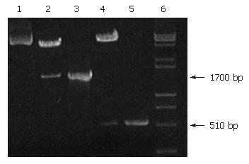
Figure 1 Agarose gel electrophoresis analysis of recombinant pIRES-ureB-IL-2.
Lane 1: pIRES after digestion with Nhe I and Xho I as a negative control, lane 2: pIRES-ureB-IL-2 after digestion with Nhe I and Xho I, lane 3: PCR product of ureB, lane 4: pIRES-ureB-IL-2 after digestion with SalI and Not I, lane 5: PCR product of IL-2, lane 6: DNA Marker (DL2000 + 15000).
Figure 2 Western blot analysis of expressed UreB protein products (A) and IL-2 protein products (B).
Lane 1: COS-7 cells transfected by pIRES-ureB, lane 2: COS-7 cells transfected by pIRES-ureB-IL-2, lane 3: COS-7 cells transfected by pIRES as a control.
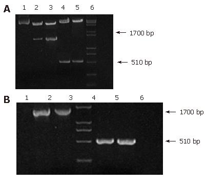
Figure 3 Agarose gel electrophoresis analysis of recombinant attenuated Salmonella typhimurium DNA vaccine strain with restriction enzyme digestion (A) (lane1: pIRES after digestion with Nhe I and Xho I, lanes 2-3: recombinant plasmid pIRES-ureB-IL-2 from strains of different generations after digestion with Nhe I and Xho I, lanes 4-5: pIRES-ureB-IL-2 from strains of different generations after digestion with Sal I and Not I, lane 6: DNA marker: (DL2000 + 15 000) and identification of recombinant attenuated Salmonella typhimurium DNA vaccine strain carrying ureB by PCR (B) (lane1: product amplified from pIRES as a negative control, Lane4: Marker (DL2000); Lanes 2-3: ureB amplified from strains of different generations, lane 4: marker-DL2000, lanes 5-6: IL-2 amplified from strains of different generations.
-
Citation: Xu C, Li ZS, Du YQ, Gong YF, Yang H, Sun B, Jin J. Construction of recombinant attenuated Salmonella typhimurium DNA vaccine expressing
H pylori ureB and IL-2. World J Gastroenterol 2007; 13(6): 939-944 - URL: https://www.wjgnet.com/1007-9327/full/v13/i6/939.htm
- DOI: https://dx.doi.org/10.3748/wjg.v13.i6.939


