Copyright
©2007 Baishideng Publishing Group Inc.
World J Gastroenterol. Dec 21, 2007; 13(47): 6441-6443
Published online Dec 21, 2007. doi: 10.3748/wjg.v13.i47.6441
Published online Dec 21, 2007. doi: 10.3748/wjg.v13.i47.6441
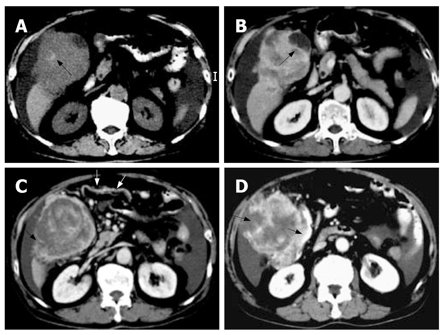
Figure 1 A 70-year-old man with hemorrhagic peritoneal malignant fibrous histiocytoma located subhepatically and presenting as massive hemoperitoneum.
A: Pre-enhanced computed tomography shows a high-density intra-lesional hemorrhage (black arrow) within the subhepatic tumor involving the anterior aspect of segment V and VI of liver. Ascites are also noted; B: Post-enhanced CT shows heterogeneous enhancement of the subhepatic mass directly invading the gallbladder (black arrow); C: Post-enhanced CT study at a lower level shows enlarged omental branches (white arrows) of the gastroepiploic artery supplying the subhepatic tumor. A cleavage plane (black arrow) between the subhepatic mass and the liver tip parenchyma was identified, suggestive of an extra-hepatic origin of the mass; D: Post-enhanced CT study at the lower tumor level reveals the large subhepatic mass directly invades the adjacent hepatic flexure of colon (thick black arrow). Tumor rupture with irregular tumor margin (thin black arrow) is also evident.
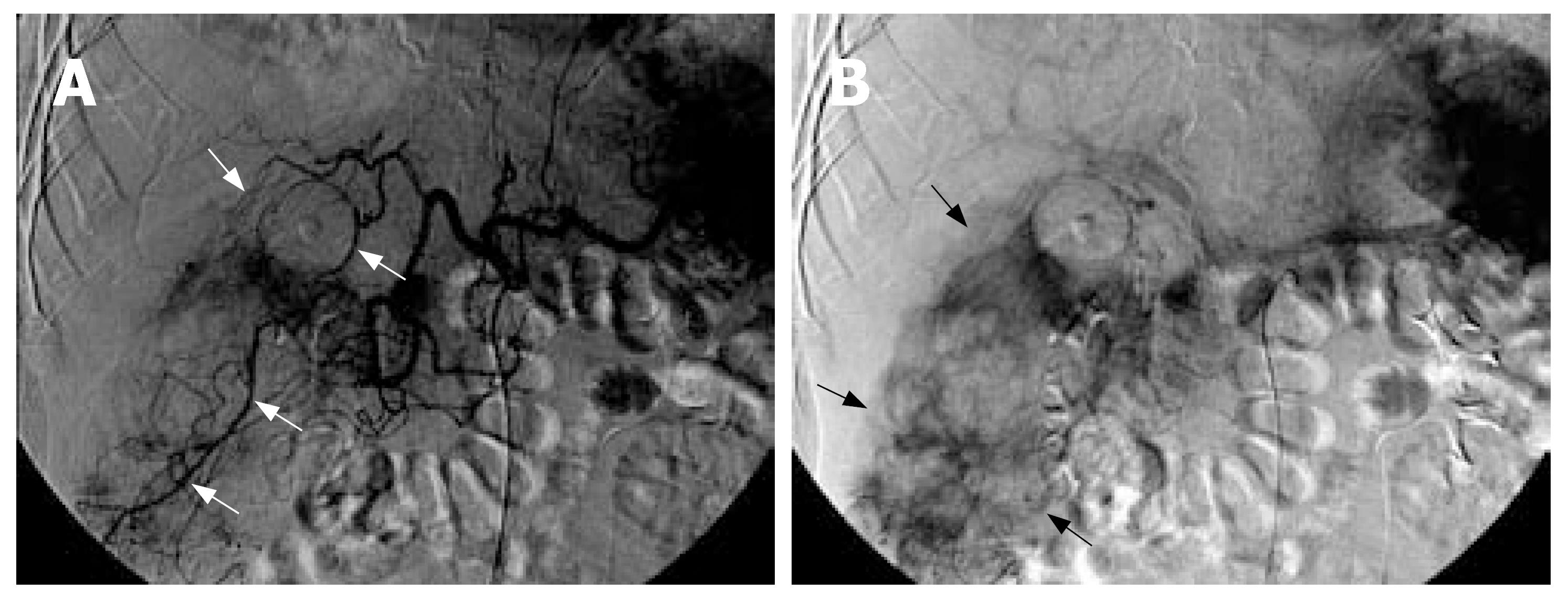
Figure 2 A: Arterial phase image of the celiac arteriogram shows hypertrophied right inferior hepatic arteries, cystic artery and omental branches of gastroepiploic artery (white arrows) supplying the hypervascular subhepatic mass; B: Venous phase shows prominent tumor stains in the peripheral portion of the huge subhepatic mass (black arrows).
- Citation: Chen HC, Chen CJ, Jeng CM, Yang CM. Malignant fibrous histiocytoma presenting as hemoperitoneum mimicking hepatocellular carcinoma rupture. World J Gastroenterol 2007; 13(47): 6441-6443
- URL: https://www.wjgnet.com/1007-9327/full/v13/i47/6441.htm
- DOI: https://dx.doi.org/10.3748/wjg.v13.i47.6441


