Copyright
©2007 Baishideng Publishing Group Co.
World J Gastroenterol. Sep 21, 2007; 13(35): 4781-4783
Published online Sep 21, 2007. doi: 10.3748/wjg.v13.i35.4781
Published online Sep 21, 2007. doi: 10.3748/wjg.v13.i35.4781
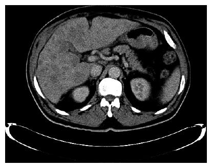
Figure 1 CT scan of the abdomen shows gastric wall thickening, several lymph nodes, and liver extensively replaced with metastatic disease.
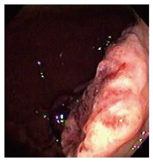
Figure 2 Upper Endoscopy shows 8-10 cm exophytic-ulcerating lesion along the greater curvature, extending from mid body to antrum
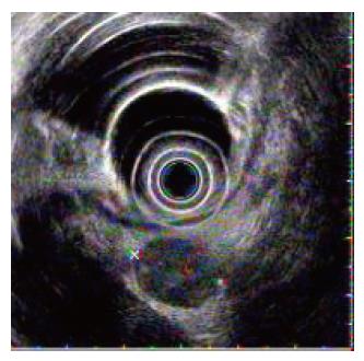
Figure 3 EUS showing a mass extending through all layers of the stomach.
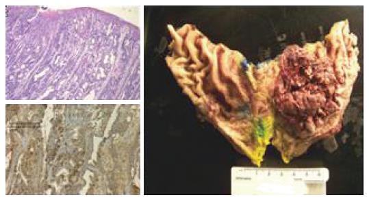
Figure 4 Immunohistochemical stains for alpha-fetoprotein strongly reactive with tumor cells.
- Citation: Singh M, Arya M, Anand S, Sandar N. Gastric adenocarcinoma with features of endodermal sinus tumor. World J Gastroenterol 2007; 13(35): 4781-4783
- URL: https://www.wjgnet.com/1007-9327/full/v13/i35/4781.htm
- DOI: https://dx.doi.org/10.3748/wjg.v13.i35.4781


