Copyright
©2007 Baishideng Publishing Group Co.
World J Gastroenterol. Apr 21, 2007; 13(15): 2243-2246
Published online Apr 21, 2007. doi: 10.3748/wjg.v13.i15.2243
Published online Apr 21, 2007. doi: 10.3748/wjg.v13.i15.2243
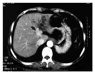
Figure 1 CT image with contrast material at the level of the upper abdomen demonstrating a well enhanced mass lesion on the left side of the hepatic hilum (arrow) along with dilated left intrahepatic bile ducts.
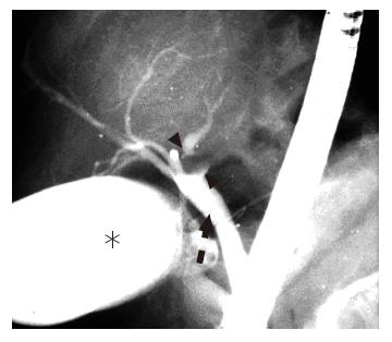
Figure 2 Endoscopic retrograde cholangiography showing a tumor in the left hepatic duct (arrow-heads) with no involvement of the right hepatic duct.
The asterisk shows the gallbladder and the dashed arrow the common bile duct.
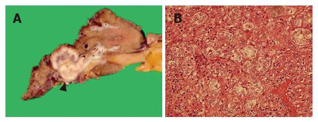
Figure 3 A tumor arising from the left hepatic duct (arrow-head) with invasion into the left intrahepatic bile ducts and the hepatic parenchyma (A) and a moderately differentiated adenocarcinoma (B) in the specimen from the patient's liver.
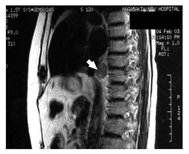
Figure 4 Magnetic resonance imaging localizes the nodule to the posterior mediastinum (arrow).
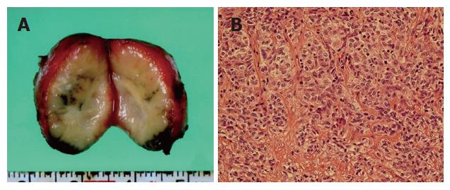
Figure 5 A well-capsulated solid mass at the cut surface (A) and a well-capsulated solid lymph node containing moderately differentiated adenocarcinoma similar in appearance to the pathology of the primary tumor (B) in the specimen of the patient's liver.
- Citation: Ito Y, Tajima Y, Fujita F, Tsutsumi R, Kuroki T, Kanematsu T. Solitary recurrence of hilar cholangiocarcinoma in a mediastinal lymph node two years after curative resection. World J Gastroenterol 2007; 13(15): 2243-2246
- URL: https://www.wjgnet.com/1007-9327/full/v13/i15/2243.htm
- DOI: https://dx.doi.org/10.3748/wjg.v13.i15.2243


