Copyright
©2007 Baishideng Publishing Group Co.
World J Gastroenterol. Apr 21, 2007; 13(15): 2243-2246
Published online Apr 21, 2007. doi: 10.3748/wjg.v13.i15.2243
Published online Apr 21, 2007. doi: 10.3748/wjg.v13.i15.2243
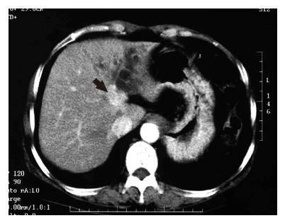
Figure 1 CT image with contrast material at the level of the upper abdomen demonstrating a well enhanced mass lesion on the left side of the hepatic hilum (arrow) along with dilated left intrahepatic bile ducts.
- Citation: Ito Y, Tajima Y, Fujita F, Tsutsumi R, Kuroki T, Kanematsu T. Solitary recurrence of hilar cholangiocarcinoma in a mediastinal lymph node two years after curative resection. World J Gastroenterol 2007; 13(15): 2243-2246
- URL: https://www.wjgnet.com/1007-9327/full/v13/i15/2243.htm
- DOI: https://dx.doi.org/10.3748/wjg.v13.i15.2243


