Copyright
©2007 Baishideng Publishing Group Co.
World J Gastroenterol. Apr 7, 2007; 13(13): 1912-1927
Published online Apr 7, 2007. doi: 10.3748/wjg.v13.i13.1912
Published online Apr 7, 2007. doi: 10.3748/wjg.v13.i13.1912
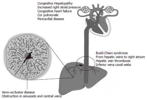
Figure 1 Site of venous obstruction in veno-occlusive disease, Budd-Chiari syndrome, and congestive hepatopathy.
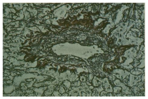
Figure 2 Histological examination demonstrates partial central vein occlusion in a patient with veno-occlusive disease.
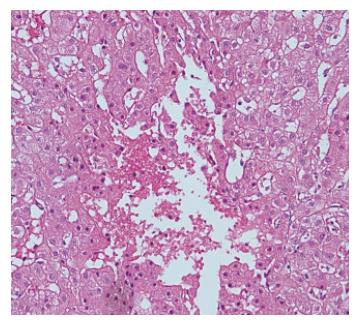
Figure 3 Histological examination shows sinusoidal dilatation and congestion.
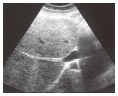
Figure 4 Abdominal ultrasonography reveals thick and obliterated middle hepatic veins.
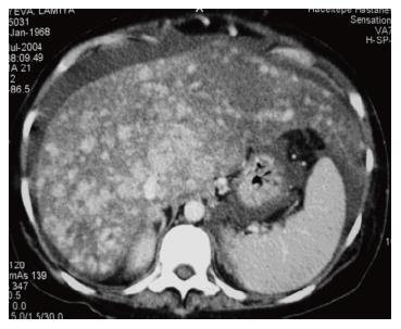
Figure 5 Computed tomography showing central contrast enhancement in the liver.
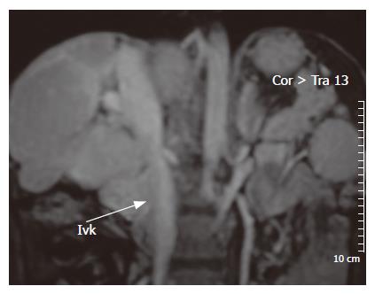
Figure 6 Magnetic resonance imaging of the liver with contrasting agent shows that inferior vena cava is compressed by hypertrophy of caudate lobe and gross lobulation of the liver.
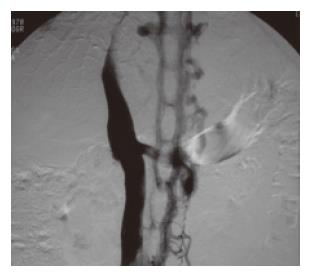
Figure 7 Almost completely obstructed IVC and shows collateral drainage through azygous system.
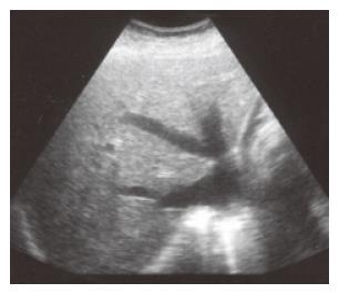
Figure 8 Ultrasound of the liver shows gross dilatation in the inferior vena cava and hepatic V.
- Citation: Bayraktar UD, Seren S, Bayraktar Y. Hepatic venous outflow obstruction: Three similar syndromes. World J Gastroenterol 2007; 13(13): 1912-1927
- URL: https://www.wjgnet.com/1007-9327/full/v13/i13/1912.htm
- DOI: https://dx.doi.org/10.3748/wjg.v13.i13.1912


