Copyright
©2006 Baishideng Publishing Group Co.
World J Gastroenterol. Aug 14, 2006; 12(30): 4922-4926
Published online Aug 14, 2006. doi: 10.3748/wjg.v12.i30.4922
Published online Aug 14, 2006. doi: 10.3748/wjg.v12.i30.4922
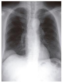
Figure 1 An extrapleural tumor projecting from the right diaphragm observed on chest roentgenography.
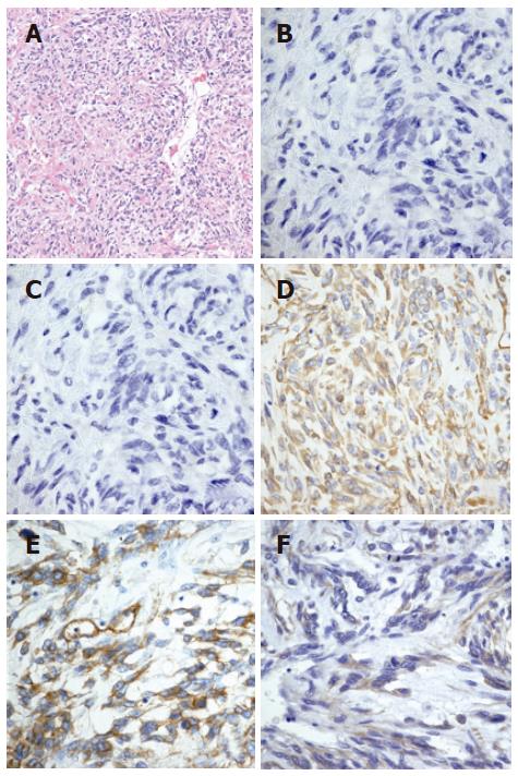
Figure 2 Spindle-shaped cells exhibiting nuclear atypicality or division together with collagen deposition (hematoxylin and eosin staining) (A), tumor cells negative for SMA (B) or CD117 (c-kit) (C) and positive for vimentin (D), CD34 (E) and CD99 (F) on histological examination.
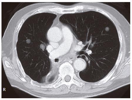
Figure 3 Multiple nodular lesions in the bilateral lung fields observed on chest computed tomography (CT).
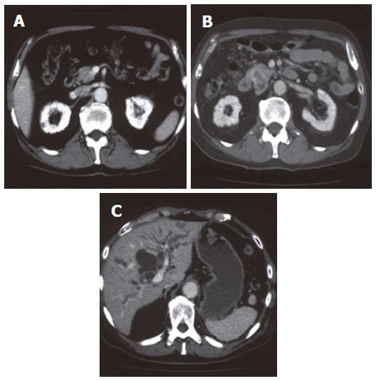
Figure 4 The pancreatic head tumor not detected by preoperative abdominal CT (A), a 25 mm × 17 mm tumor in the pancreatic head detected by abdominal CT with its periphery enhanced by contrast material in the delayed phase (B) and dilatation of intra- and extra-hepatic bile ducts observed (C).
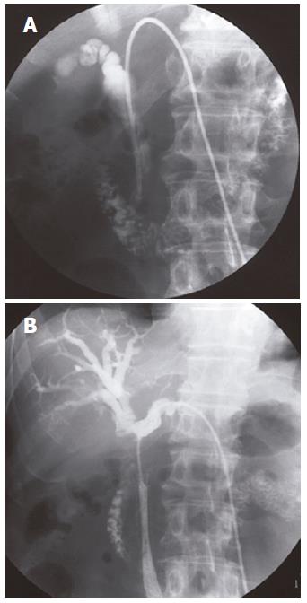
Figure 5 A marked stenosis of the inferior part of the CBD observed on cholangiography via a PTBD tube (A), an obstruction from inferior part of CBD to the confluence of the right and left extrahepatic bile ducts involving the whole stent on reperformed cholangiography (B) on microscopic examination.
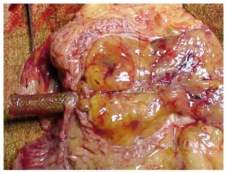
Figure 6 Macroscopic findings on autopsy.
A dissected (120 mm × 30 mm × 20 mm) tumor spreading from the pancreatic head to the hepatic hilum and involving bilateral hepatic ducts. White arrow shows the pancreatic head tumor and black arrow shows the metallic stent.
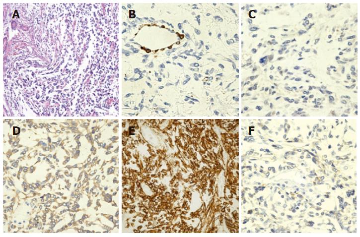
Figure 7 Spindle-shaped cells exhibiting nuclear atypicality or division together with collagen deposition (hematoxylin and eosin staining) (A), tumor cells negative for SMA (B) or CD117 (c-kit) (C) and positive for vimentin (D), CD34 (E) and CD99 (F) on histological examination.
These findings are consistent with those on malignant solitary fibrous tumor of the pleura, resected in July 2002.
- Citation: Yamada N, Okuse C, Nomoto M, Orita M, Katakura Y, Ishii T, Shinmyo T, Osada H, Maeda I, Yotsuyanagi H, Suzuki M, Itoh F. Obstructive jaundice caused by secondary pancreatic tumor from malignant solitary fibrous tumor of pleura: A case report. World J Gastroenterol 2006; 12(30): 4922-4926
- URL: https://www.wjgnet.com/1007-9327/full/v12/i30/4922.htm
- DOI: https://dx.doi.org/10.3748/wjg.v12.i30.4922


