
















©2006 Baishideng Publishing Group Co.
World J Gastroenterol. Aug 14, 2006; 12(30): 4922-4926
Published online Aug 14, 2006. doi: 10.3748/wjg.v12.i30.4922
Published online Aug 14, 2006. doi: 10.3748/wjg.v12.i30.4922
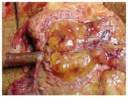
Figure 6 Macroscopic findings on autopsy.
A dissected (120 mm × 30 mm × 20 mm) tumor spreading from the pancreatic head to the hepatic hilum and involving bilateral hepatic ducts. White arrow shows the pancreatic head tumor and black arrow shows the metallic stent.
- Citation: Yamada N, Okuse C, Nomoto M, Orita M, Katakura Y, Ishii T, Shinmyo T, Osada H, Maeda I, Yotsuyanagi H, Suzuki M, Itoh F. Obstructive jaundice caused by secondary pancreatic tumor from malignant solitary fibrous tumor of pleura: A case report. World J Gastroenterol 2006; 12(30): 4922-4926
- URL: https://www.wjgnet.com/1007-9327/full/v12/i30/4922.htm
- DOI: https://dx.doi.org/10.3748/wjg.v12.i30.4922