Copyright
©2006 Baishideng Publishing Group Co.
World J Gastroenterol. Aug 14, 2006; 12(30): 4908-4910
Published online Aug 14, 2006. doi: 10.3748/wjg.v12.i30.4908
Published online Aug 14, 2006. doi: 10.3748/wjg.v12.i30.4908
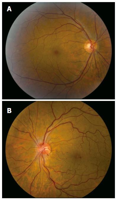
Figure 1 Normal fundus in the right eye (A) and marked venous tortuosity and scattered retinal hemorrhages in the left eye (B).
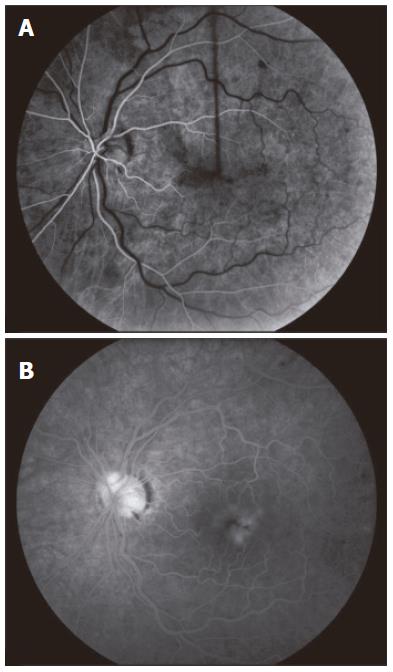
Figure 2 Left eye angiogram.
A: Early phase image revealing delayed venous filling; B: Late phase image revealing leakage of fluorescein in the macula.
- Citation: Zandieh I, Adenwalla M, Cheong-Lee C, Ma PE, Yoshida EM. Retinal vein thrombosis associated with pegylated-interferon and ribavirin combination therapy for chronic hepatitis C. World J Gastroenterol 2006; 12(30): 4908-4910
- URL: https://www.wjgnet.com/1007-9327/full/v12/i30/4908.htm
- DOI: https://dx.doi.org/10.3748/wjg.v12.i30.4908


