Copyright
©2006 Baishideng Publishing Group Co.
World J Gastroenterol. Jan 14, 2006; 12(2): 228-233
Published online Jan 14, 2006. doi: 10.3748/wjg.v12.i2.228
Published online Jan 14, 2006. doi: 10.3748/wjg.v12.i2.228
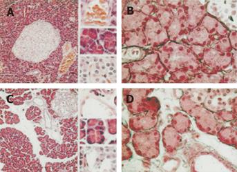
Figure 1 Pancreatic tissues from sham and BDL-60 rats were stained with hematoxylin and eosin (panels A and C) and Gomori (panels B and D).
In panels A and C, magnification is 100x and 400x (upper: small vessel; middle: acini; lower: Langerhans islet) and in panels B and D, magnification is 400x. The extracellular matrix was enlarged in pancreas isolated from BDL-60 rats and most of the enlarged extracellular matrix consisted of edema. No increase in reticulin fibers was observed with the Gomori technique. n = 3 in each group.
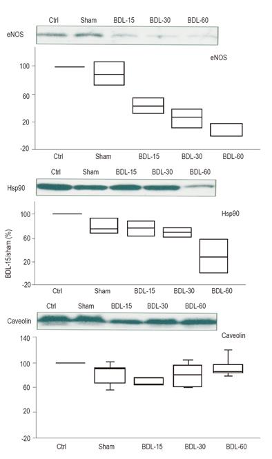
Figure 2 Endothelial nitric oxide synthase (eNOS), heat shock protein 90(Hsp90),
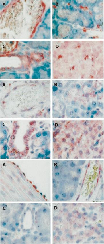
Figure 3 Immunostaining of caveolin (I) in pancreas isolated from BDL-30 rat.
Caveolin was detected in epithelial cells lining the pancreatic ductules (C), cells within the islets of Langerhans (D), and in the apex of acinar cells (B). Cavolin was also present in endothelial lining cells in veinules (A), microvessels surrounding acini (B), and microvessels inside Langerhans islets (D). Immunostaining of Hsp90 (II) in pancreas isolated from BDL-15 rat. Hsp90 was present in epithelial cells lining the pancreatic ductules (C), cells within the islets of Langerhans (D), and in the apex of acinar cells (B). Hsp90 was also present in endothelial lining cells in veinules (A). Immunostaining of endothelial nitric oxide synthase (III) in pancreas isolated from BDL-15 rat. The protein was present in endothelial lining cells (B) and cells within the islets of Langerhans (D). In aorta isolated from BDL-60 rats (A), eNOS is more heavily stained. Magnification is 400x.
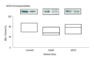
Figure 4 Endothelial NO synthase (eNOS) immunoprecipitation from pancreatic extracts.
Immunoprecipitates were Western blotted with Hsp90, caveolin, and eNOS antibodies. Pancreatic proteins were collected from sham rats and rats which had a 15 days bile duct ligation (BDL-15).
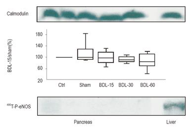
Figure 5 Calmodulin and 495Thr-P-eNOS expression in pancreatic lysates isolated from control (Ctrl), sham rats, and rats with bile duct ligation (BDL) 15, 30, and 60 days before tissue collection.
Densitometry was measured in ≥ 4 rats in each group. Liver homogenate serves as control.
- Citation: Frossard JL, Quadri R, Hadengue A, Morel P, Pastor CM. Endothelial nitric oxide synthase regulation is altered in pancreas from cirrhotic rats. World J Gastroenterol 2006; 12(2): 228-233
- URL: https://www.wjgnet.com/1007-9327/full/v12/i2/228.htm
- DOI: https://dx.doi.org/10.3748/wjg.v12.i2.228


