Copyright
©2006 Baishideng Publishing Group Co.
World J Gastroenterol. Apr 7, 2006; 12(13): 2060-2064
Published online Apr 7, 2006. doi: 10.3748/wjg.v12.i13.2060
Published online Apr 7, 2006. doi: 10.3748/wjg.v12.i13.2060
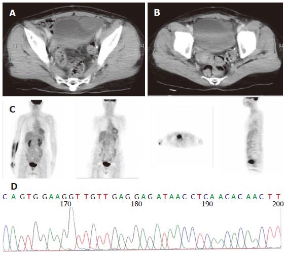
Figure 1 (A) Abdominal CT showing a tumor located near the urinary bladder (arrow); (B) abdominal CT showing complete response without tumor at the same level as Figure 1A; (C) [18F] fluoro-2-deoxy-D-glucose positron-emission tomography (PET) scanning PET revealing no tumor with metabolic activity in the whole body; (D) Direct sequencing analysis of DNA from patient 1 showed deletion and insertion mutation at codons 563-572 in exon 11 (arrow).
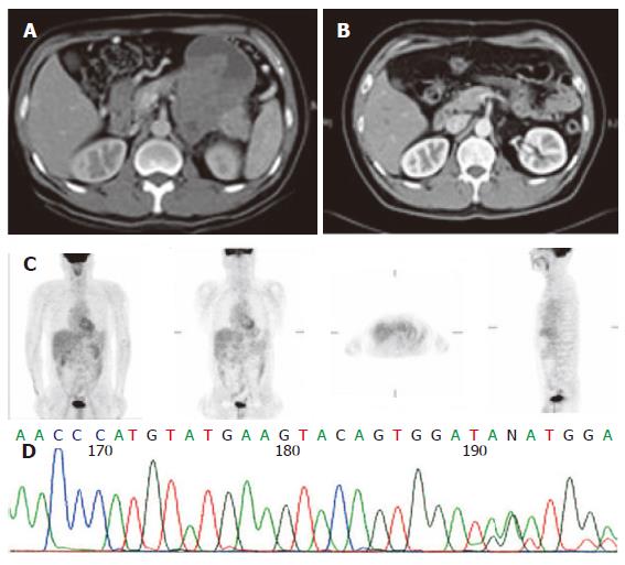
Figure 2 (A) Abdominal CT showing a huge retroperitoneal tumor invading the pancreas (arrow); (B) abdominal CT showing complete response without tumor at the same level as Figure 2A; (C) PET showing no tumor with metabolic activity in the whole body; (D) direct sequencing analysis of DNA from patient 2 showed deletion and insertion mutation at codons 556-557 in exon 11 (arrow).
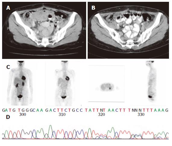
Figure 3 (A) Abdominal CT showing a tumor located near the ileum (arrow); (B) abdominal CT revealing complete response without tumor at the same level as Figure 3A; (C) PET showing no tumor with metabolic activity in the whole body; (D) direct sequencing analysis of DNA from patient 3 showed insertion AY at codons 502-503 in exon 9 (arrow).
- Citation: Chiang KC, Chen TW, Yeh CN, Liu FY, Lee HL, Jan YY. Advanced gastrointestinal stromal tumor patients with complete response after treatment with imatinib mesylate. World J Gastroenterol 2006; 12(13): 2060-2064
- URL: https://www.wjgnet.com/1007-9327/full/v12/i13/2060.htm
- DOI: https://dx.doi.org/10.3748/wjg.v12.i13.2060


