Copyright
©2005 Baishideng Publishing Group Inc.
World J Gastroenterol. Feb 14, 2005; 11(6): 875-879
Published online Feb 14, 2005. doi: 10.3748/wjg.v11.i6.875
Published online Feb 14, 2005. doi: 10.3748/wjg.v11.i6.875
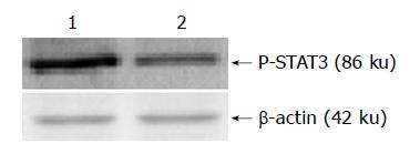
Figure 1 Western blot analysis of phospho-STAT3 in different cell lines.
Lane 1: SGC7901 cell line; lane 2: SGC7901/R cell line.
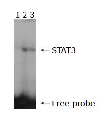
Figure 2 EMSA analysis of STAT3 DNA-binding activity in different cell lines.
Lane 1: Competitive probe; lane 2: SGC7901/R cell line; lane 3: SGC7901 cell line.
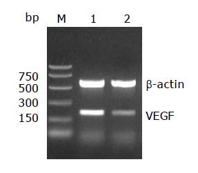
Figure 3 Identification of VEGF mRNA by RT-PCR in different cell lines.
M: Ladder marker; lane 1: SGC7901 cell line; lane 2: SGC7901/R cell line.
Figure 4 Immunocytochemical staining of VEGF in two human stomach adenocarcinoma cell lines (DAB×400).
A: parental cell line SGC7901; B: drug-resistant cell line SGC7901/R.
- Citation: Yu LF, Cheng Y, Qiao MM, Zhang YP, Wu YL. Activation of STAT3 signaling in human stomach adenocarcinoma drug-resistant cell line and its relationship with expression of vascular endothelial growth factor. World J Gastroenterol 2005; 11(6): 875-879
- URL: https://www.wjgnet.com/1007-9327/full/v11/i6/875.htm
- DOI: https://dx.doi.org/10.3748/wjg.v11.i6.875


