Copyright
©2005 Baishideng Publishing Group Inc.
World J Gastroenterol. Feb 7, 2005; 11(5): 726-728
Published online Feb 7, 2005. doi: 10.3748/wjg.v11.i5.726
Published online Feb 7, 2005. doi: 10.3748/wjg.v11.i5.726
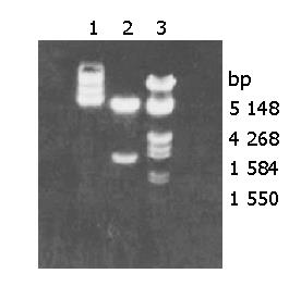
Figure 1 Macrorestriction map of mutated IκBα Plasmi.
Lane 1: Plasmid DNA; lane 2: Plasmid DNA cut by Xbal and HandIII; lane 3: DNA marker.
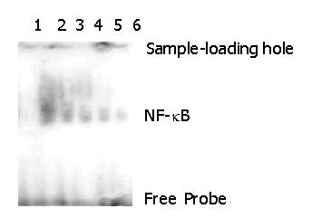
Figure 2 EMSA of QBC939 and QBC939HCVC +cells transfected with mutated IκBα plasmid.
Lane 1: Control group; lane 2: QBC939HCVC +cells without transfected with mutated IκBα; lane 3: QBC939 cells not transfected with mutated IκBα; lanes 4 and 5: QBC939HCVC + cells transfected with mutated IκBα; lane 6: QBC939 cells transfected with mutated IκBα.
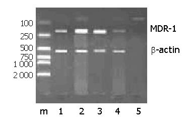
Figure 3 Expression of MDR-1mRNA in the QBC939 cells transfected with mutated IκBα.
M: DNA marker. Lane 1: The QBC939HCVC+ cells transfected with mutated IκBα, lane 2: The QBC939HCVC + cells not transfected with mutated IκBα; lane 3: The QBC939 cells not transfected with mutated IκBα; lane 4: The QBC939 cells transfected with mutated IκBα.
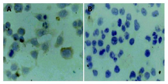
Figure 4 Expression intensity of P-GP protein in QBC939 and QBC939HCVC+ cells transfected with mutated IκBα.
A: Expression of P-GP in the non-transfection Group, LSAB ×400; B: Expression of P-GP after transfection of mutated IκBα, LSAB ×400.
- Citation: Chen RF, Li ZH, Kong XH, Chen JS. Effect of mutated IκBα transfection on multidrug resistance in hilar cholangiocarcinoma cell lines. World J Gastroenterol 2005; 11(5): 726-728
- URL: https://www.wjgnet.com/1007-9327/full/v11/i5/726.htm
- DOI: https://dx.doi.org/10.3748/wjg.v11.i5.726


