Copyright
©2005 Baishideng Publishing Group Inc.
World J Gastroenterol. Dec 28, 2005; 11(48): 7597-7601
Published online Dec 28, 2005. doi: 10.3748/wjg.v11.i48.7597
Published online Dec 28, 2005. doi: 10.3748/wjg.v11.i48.7597
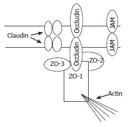
Figure 1 Tight junction (TJ) structure with emphasis on proteins of the TJ membrane domain: ZO family members and their interaction with actin filaments.
Figure 2 Phase-contrast micrographs (10×) showing control (A) and gliadin-treated multicellular tumor spheroids of the human colon adenocarcinoma LoVo cell line (B) after 11 d of culture; Scanning electron micrographs: showing the ovoid control spheroids (C) and their very compact, densely organized and tightly packed structure that they can be clearly distinguished from each other; The surface of gliadin-treated spheroids (D) focally interrupted by irregularly distributed holes, and loss of their structural thickness and organization (E).
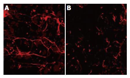
Figure 3 Confocal laser scanning micrograph in which TRICT-phalloidin highlights the organization of F-actin in multicellular tumor spheroids.
The control spheroids (A) have a "chicken-wire" distribution under the plasma membrane, whereas the treated spheroids (B) show a reorganized actin cytoskeleton.
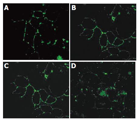
Figure 4 Confocal microscopy immunolocalization of occludin and zonula occluden-1 (ZO-1) in LoVo multicellular tumor spheroids.
In the control spheroids, occludin and ZO-1 localized sharply in the apical region of the lateral membrane are visualized en face as a ring pattern (A and C), whereas the distribution of occludin and ZO-1 in the spheroids exposed to gliadin for 4 d is far from the lateral TJ membrane (B and D).
-
Citation: Dolfini E, Roncoroni L, Elli L, Fumagalli C, Colombo R, Ramponi S, Forlani F, Bardella MT. Cytoskeleton reorganization and ultrastructural damage induced by gliadin in a three-dimensional
in vitro model. World J Gastroenterol 2005; 11(48): 7597-7601 - URL: https://www.wjgnet.com/1007-9327/full/v11/i48/7597.htm
- DOI: https://dx.doi.org/10.3748/wjg.v11.i48.7597


