Copyright
©2005 Baishideng Publishing Group Inc.
World J Gastroenterol. Nov 28, 2005; 11(44): 6954-6959
Published online Nov 28, 2005. doi: 10.3748/wjg.v11.i44.6954
Published online Nov 28, 2005. doi: 10.3748/wjg.v11.i44.6954
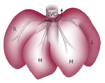
Figure 1 Anterior view of left tri-segmentectomy.
Bold line indicates the left and middle hepatic veins ligated by transfixing suture; arrow indicates parenchymal transection of the left lobe and right paramedian lobe; L: left lateral lobe; M: median lobe; R: right lateral lobe; IVC: inferior vena cava.
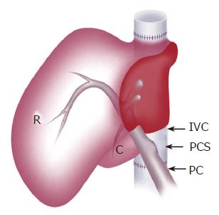
Figure 2 Schematic view of the liver graft with PCS placed by side-to-side anastomosis of PV and IVC.
R: right lateral lobe; C: caudate lobe; PV: portal vein; IVC: inferior vena cava; PCS: portocaval shunt.
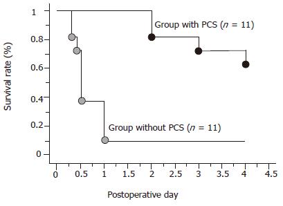
Figure 3 Survival rates.
bP<0.01 vs the group without PCS.
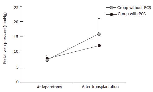
Figure 4 Changes of portal vein pressure (n = 11).
aP<0.05 vs the group with PCS.
Figure 5 Histological findings of hepatic tissues after reperfusion (×100).
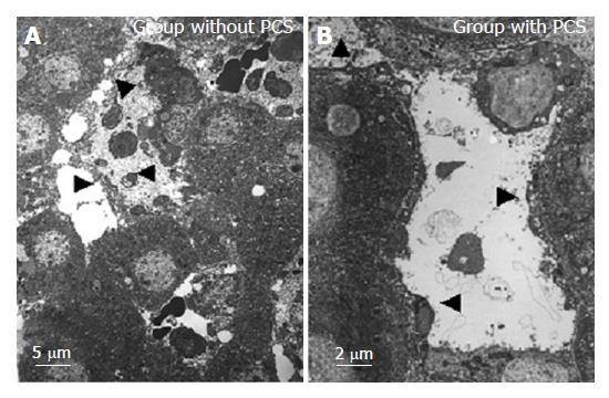
Figure 6 Transmission electron microscopical findings of the sinusoid after reperfusion.
Arrowheads indicate the destroyed or preserved or detached sinusoidal endothelial cells into the sinusoidal space with destructed or intact Disse’s spaces.
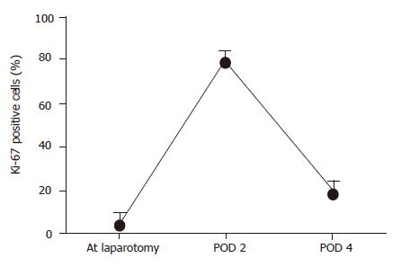
Figure 7 Ki-67 detection in the pigs that survived for more than 4 d in the group with PCS (n = 8).
- Citation: Wang HS, Ohkohchi N, Enomoto Y, Usuda M, Miyagi S, Asakura T, Masuoka H, Aiso T, Fukushima K, Narita T, Yamaya H, Nakamura A, Sekiguchi S, Kawagishi N, Sato A, Satomi S. Excessive portal flow causes graft failure in extremely small-for-size liver transplantation in pigs. World J Gastroenterol 2005; 11(44): 6954-6959
- URL: https://www.wjgnet.com/1007-9327/full/v11/i44/6954.htm
- DOI: https://dx.doi.org/10.3748/wjg.v11.i44.6954


