
















©2005 Baishideng Publishing Group Inc.
World J Gastroenterol. Nov 28, 2005; 11(44): 6954-6959
Published online Nov 28, 2005. doi: 10.3748/wjg.v11.i44.6954
Published online Nov 28, 2005. doi: 10.3748/wjg.v11.i44.6954
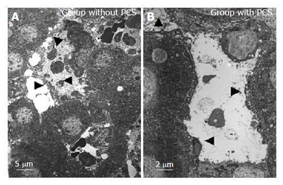
Figure 6 Transmission electron microscopical findings of the sinusoid after reperfusion.
Arrowheads indicate the destroyed or preserved or detached sinusoidal endothelial cells into the sinusoidal space with destructed or intact Disse’s spaces.
- Citation: Wang HS, Ohkohchi N, Enomoto Y, Usuda M, Miyagi S, Asakura T, Masuoka H, Aiso T, Fukushima K, Narita T, Yamaya H, Nakamura A, Sekiguchi S, Kawagishi N, Sato A, Satomi S. Excessive portal flow causes graft failure in extremely small-for-size liver transplantation in pigs. World J Gastroenterol 2005; 11(44): 6954-6959
- URL: https://www.wjgnet.com/1007-9327/full/v11/i44/6954.htm
- DOI: https://dx.doi.org/10.3748/wjg.v11.i44.6954