Copyright
©2005 Baishideng Publishing Group Inc.
World J Gastroenterol. Jan 28, 2005; 11(4): 614-615
Published online Jan 28, 2005. doi: 10.3748/wjg.v11.i4.614
Published online Jan 28, 2005. doi: 10.3748/wjg.v11.i4.614
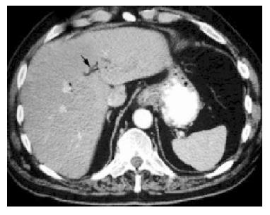
Figure 1 Air within the intrahepatic bile ducts [arrow] during the first CT scan.
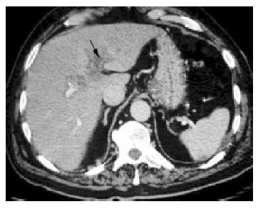
Figure 2 Thrombosis within the portal vein [large arrow] during the second CT scan.
A splenic infarct is also seen [small arrow].
- Citation: Wireko M, Berry PA, Brennan J, Aga R. Unrecognized pylephlebitis causing life-threatening septic shock: A case report. World J Gastroenterol 2005; 11(4): 614-615
- URL: https://www.wjgnet.com/1007-9327/full/v11/i4/614.htm
- DOI: https://dx.doi.org/10.3748/wjg.v11.i4.614


