Copyright
©2005 Baishideng Publishing Group Inc.
World J Gastroenterol. Sep 21, 2005; 11(35): 5455-5459
Published online Sep 21, 2005. doi: 10.3748/wjg.v11.i35.5455
Published online Sep 21, 2005. doi: 10.3748/wjg.v11.i35.5455
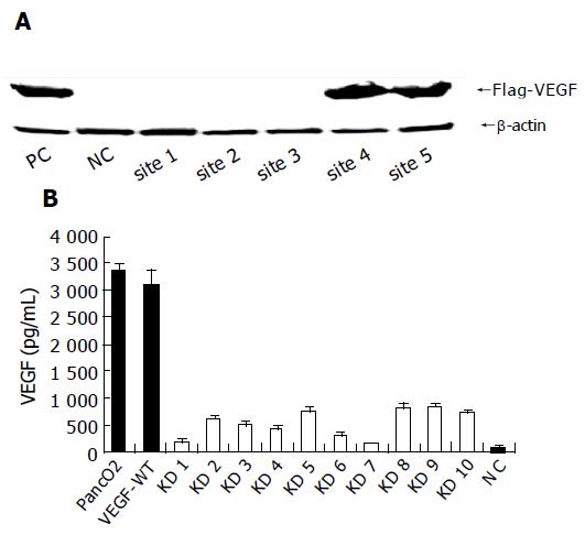
Figure 1 Establishment of VEGF knockdown pancreatic cancer cell lines.
A: Western blot showing marked reduction of VEGF expression in knockdown cells. Screening of the most effective targeting site for the VEGF gene. B: VEGF production of stable knockdown PancO2 cell clones. VEGF-WT and VEGF-KD clones (KD 1-10) were grown in 6-well plates, and the VEGF concentration in culture supernatant was measured using ELISA in triplicates. The growth medium was used as negative control (NC).
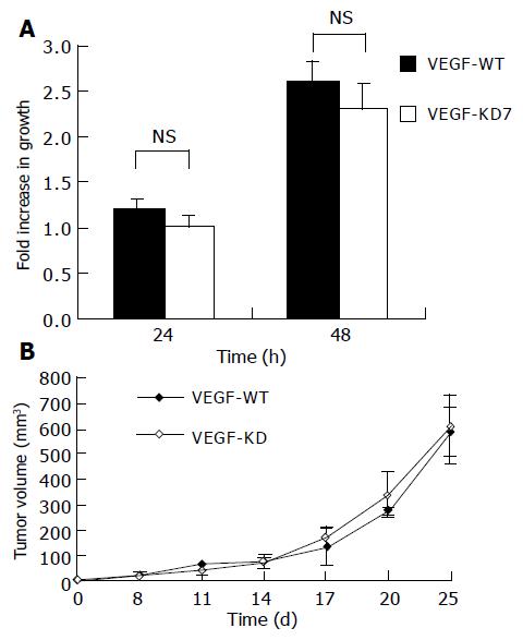
Figure 2 Effect of VEGF knockdown on cell proliferation.
A: 3×10⁴ VEGF-KD7 or VEGF-WT cells were grown in 24-well plates, and the numbers of viable cells were assessed using the MTT assay at 24 (P = 0.082) and 48 h (P = 0.078) (n = 4, mean±SD); B: 2×10⁶ VEGF-KD7 or VEGF-WT cells were injected into C57BL/6 mice subicutaneous, the tumor size was measured, and the volume was calculated as [(length (mm)×width (mm)²)]/2 (n = 4, P = 0.18, mean±SD).
Figure 3 Effect of VEGF knockdown in a tumor grafted mouse ascites model.
A: The ascites volume of mice inoculated with VEGF-KD7 or VEGF-WT cells (n = 5, P = 0.10, mean±SD); B: The VEGF concentration in ascites. VEGF in the ascites was measured using ELISA (n = 4, P = 0.054, mean±SD). C: 1106 VEGF-KD7 or VEGF-WT cells were inoculated into mice intraperitoneally and the mice were weighed (n = 8, P≥0.05 in each data point).
- Citation: Guleng B, Tateishi K, Kanai F, Jazag A, Ohta M, Asaoka Y, Ijichi H, Tanaka Y, Imamura J, Ikenoue T, Fukushima Y, Morikane K, Miyagishi M, Taira K, Kawabe T, Omata M. Cancer-derived VEGF plays no role in malignant ascites formation in the mouse. World J Gastroenterol 2005; 11(35): 5455-5459
- URL: https://www.wjgnet.com/1007-9327/full/v11/i35/5455.htm
- DOI: https://dx.doi.org/10.3748/wjg.v11.i35.5455


