Copyright
©The Author(s) 2005.
World J Gastroenterol. Aug 21, 2005; 11(31): 4899-4903
Published online Aug 21, 2005. doi: 10.3748/wjg.v11.i31.4899
Published online Aug 21, 2005. doi: 10.3748/wjg.v11.i31.4899
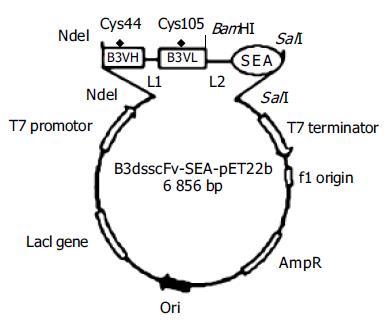
Figure 1 Schematic representation of B3scdsFv-SEA-pET22b expression plasmid.
L: polypeptide linker; L1: (GlyGlyGlyGlySer)3; L2: (GlyGlyGlySer-GlyGlySer).
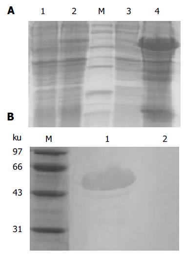
Figure 2 The SDS-PAGE analysis of expression products (A) and Western blot of inclusion body (B).
A: Lane 1: B3scdsFv-SEA- pET22b before induction; lane 2: total protein after induction; lane 3: ultrasonic supernatant of B3scdsFv-SEA-pET22b induced by 1 mmol/L IPTG; lane 4: ultrasonic deposit after induction; lane M: low molecular weight protein marker; B: Lane 1: Western blot of B3scdsFv-SEA; lane 2: negative control (total protein before induction); lane M: low molecular weight protein marker.
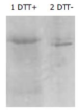
Figure 3 Gel mobility of B3scdsFv-SEA under reducing (lane 1) and non-reducing condition (lane 2).
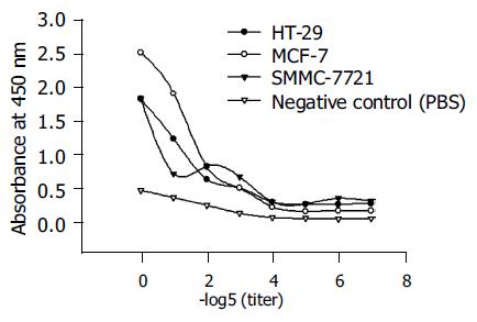
Figure 4 Binding assay of B3scdsFv-SEA to B3 antigen positive carcinoma cell lines.
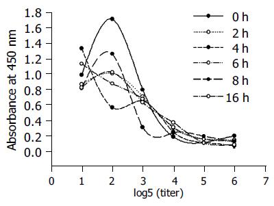
Figure 5 Stability of B3scdsFv-SEA.
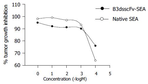
Figure 6 Cytotoxic effects of B3scdsFv-SEA on HT-29 cell line.
- Citation: Wang JL, Zheng YL, Ma R, Wang BL, Guo AG, Jiang YQ. Disulfide-stabilized single-chain antibody-targeted superantigen: Construction of a prokaryotic expression system and its functional analysis. World J Gastroenterol 2005; 11(31): 4899-4903
- URL: https://www.wjgnet.com/1007-9327/full/v11/i31/4899.htm
- DOI: https://dx.doi.org/10.3748/wjg.v11.i31.4899


