Copyright
©The Author(s) 2005.
World J Gastroenterol. Aug 14, 2005; 11(30): 4638-4643
Published online Aug 14, 2005. doi: 10.3748/wjg.v11.i30.4638
Published online Aug 14, 2005. doi: 10.3748/wjg.v11.i30.4638
Figure 1 Immunohistochemical expression of COX-2 in cirrhotic liver area (A), well- and moderately-differentiated HCC areas (B), well-differentiated HCC with trabecular arrangement and poorly-differentiated HCC with loose cohesive pattern (C) (×250).
Figure 2 Immunohistochemical staining with anti-CD68 highlights the number of histiocytes in cirrhotic liver cells (A), well-differentiated HCC (B) and poorly differentiated HCC (C).
(A, C ×250; B ×400).
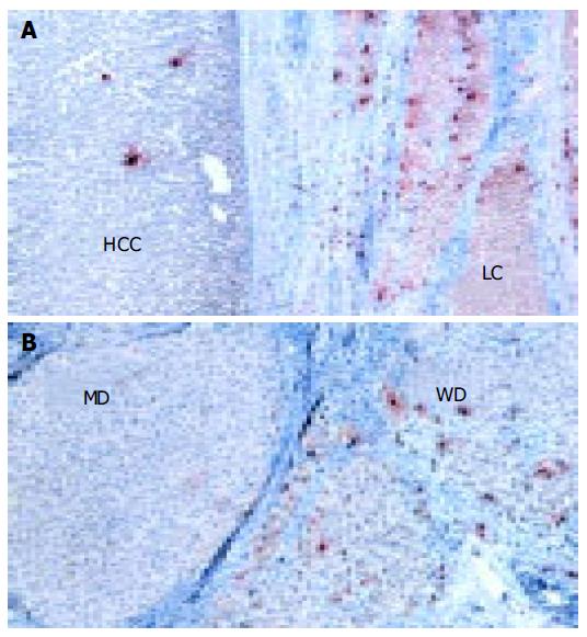
Figure 3 Immunohistochemical staining with anti-human mast cell tryptase in cirrhotic and HCC tissues (A) and in well-differentiated HCC and moderately-differentiated HCC (B).
(x 250).
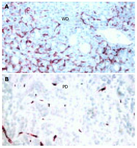
Figure 4 Immunohistochemical expression of CD34 underlining striking differences between well-differentiated HCC (A) and poorly-differentiated HCC (B).
(×250).
- Citation: Cervello M, Foderà D, Florena AM, Soresi M, Tripodo C, D’Alessandro N, Montalto G. Correlation between expression of cyclooxygenase-2 and the presence of inflammatory cells in human primary hepatocellular carcinoma: Possible role in tumor promotion and angiogenesis. World J Gastroenterol 2005; 11(30): 4638-4643
- URL: https://www.wjgnet.com/1007-9327/full/v11/i30/4638.htm
- DOI: https://dx.doi.org/10.3748/wjg.v11.i30.4638


