Copyright
©2005 Baishideng Publishing Group Inc.
World J Gastroenterol. May 28, 2005; 11(20): 3065-3069
Published online May 28, 2005. doi: 10.3748/wjg.v11.i20.3065
Published online May 28, 2005. doi: 10.3748/wjg.v11.i20.3065
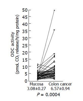
Figure 1 ODC activity in colon cancer tissues and adjacent normal tissues.
ODC activities in colon cancer tissues and adjacent normal tissues were assayed as described in Materials and methods, and the data are shown as mean±SD (pmol CO2 release/h/mg protein).
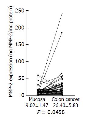
Figure 2 MMP-2 expression levels in colon cancer tissues and adjacent normal tissues.
MMP-2 expression levels in colon cancer tissues and adjacent normal tissues were analyzed by MMP-2 ELISA, and the data are shown as mean±SD (ng MMP-2/mg protein).
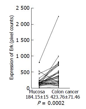
Figure 3 Erk expression in colon cancer tissues and adjacent normal tissues.
Expression of phosphorylated Erk1/2 in colon cancer tissues and adjacent normal tissues was analyzed by Western blotting using a specific antibody against phosphorylated Erk1/2, quantitated using a NIH image, and shown as mean±SD (pixel counts).
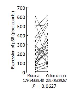
Figure 4 p38 MAP kinase expression in colon cancer tissues and adjacent normal tissues.
Expression of phosphorylated p38 MAP kinase in colon cancer tissues and adjacent normal tissues was analyzed by Western blotting using a specific antibody against phosphorylated p38 MAP kinase, quantitated using a NIH image, and are shown as mean±SD (pixel counts).
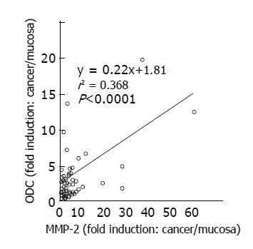
Figure 5 Relationship between ODC activities and MMP-2 expression in colon cancer tissues.
Correlation between ODC activities and MMP-2 expression levels in cancer tissues, compared to those in adjacent normal tissues was shown. Statistical significance was analyzed as described in Materials and methods.
Figure 6 Relationship between ODC activities and Erk expression in colon cancer tissues.
Correlation between ODC activities and Erk1/2 expression levels in cancer tissues, compared to those in adjacent normal tissues was shown. Statistical significance was analyzed as described in Materials and methods.
- Citation: Nemoto T, Kubota S, Ishida H, Murata N, Hashimoto D. Ornithine decarboxylase, mitogen-activated protein kinase and matrix metalloproteinase-2 expressions in human colon tumors. World J Gastroenterol 2005; 11(20): 3065-3069
- URL: https://www.wjgnet.com/1007-9327/full/v11/i20/3065.htm
- DOI: https://dx.doi.org/10.3748/wjg.v11.i20.3065


