Copyright
©2005 Baishideng Publishing Group Co.
World J Gastroenterol. Jan 14, 2005; 11(2): 296-298
Published online Jan 14, 2005. doi: 10.3748/wjg.v11.i2.296
Published online Jan 14, 2005. doi: 10.3748/wjg.v11.i2.296
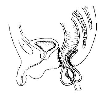
Figure 1 Transrectal dilator tied around ganglionic bowel at transition zone, and pulled through rectum.
Bowel was everted until mucocutaneous line was exposed posteriorly, but not anteriorly.
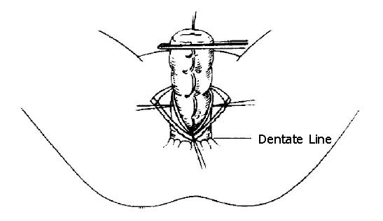
Figure 2 Ganglionic bowel exposed by resection of most dilated bowel.
The posterior wall of the aganglionic anorectum was longitudinally split in posterior wall of anorectal canal to dentate line.
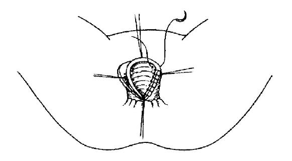
Figure 3 Suturing of seromuscular coats of rectum and colon.
The bowel was everted and exteriorized out of the anus.
- Citation: Wang G, Sun XY, Wei MF, Weng YZ. Heart-shaped anastomosis for Hirschsprung’s disease: Operative technique and long-term follow-up. World J Gastroenterol 2005; 11(2): 296-298
- URL: https://www.wjgnet.com/1007-9327/full/v11/i2/296.htm
- DOI: https://dx.doi.org/10.3748/wjg.v11.i2.296


