Copyright
©2005 Baishideng Publishing Group Inc.
World J Gastroenterol. May 21, 2005; 11(19): 2960-2966
Published online May 21, 2005. doi: 10.3748/wjg.v11.i19.2960
Published online May 21, 2005. doi: 10.3748/wjg.v11.i19.2960
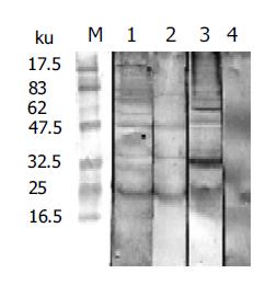
Figure 1 Immunological recognition bands of AWA in rat sera.
M: Molecular weight standards; 1: IRS; 2: NRS; 3: IMS; 4: NMS.
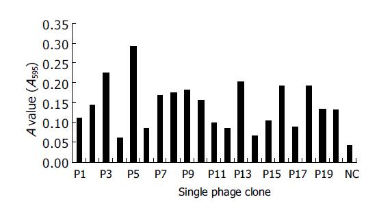
Figure 2 Detection of positive phage clones by ELISA.
P1-P20: Twenty phage clones; NC: normal controls (original peptides).
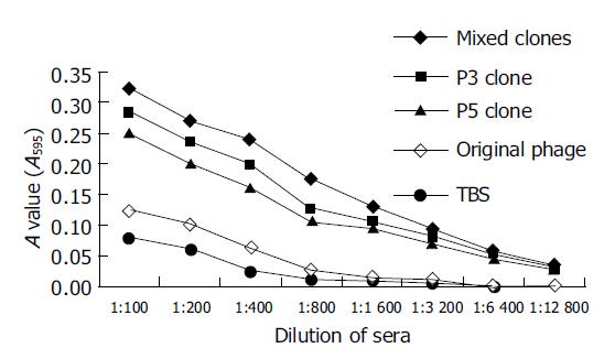
Figure 3 Antibody titer in sera of mice after the third immunization.
- Citation: Wang M, Yi XY, Li XP, Zhou DM, Larry M, Zeng XF. Phage displaying peptides mimic schistosoma antigenic epitopes selected by rat natural antibodies and protective immunity induced by their immunization in mice. World J Gastroenterol 2005; 11(19): 2960-2966
- URL: https://www.wjgnet.com/1007-9327/full/v11/i19/2960.htm
- DOI: https://dx.doi.org/10.3748/wjg.v11.i19.2960


