Copyright
©The Author(s) 2004.
World J Gastroenterol. Sep 15, 2004; 10(18): 2666-2669
Published online Sep 15, 2004. doi: 10.3748/wjg.v10.i18.2666
Published online Sep 15, 2004. doi: 10.3748/wjg.v10.i18.2666
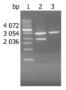
Figure 1 Detection of EcoR I-digested pBR325 and recovered long fragment by 10 g/L agarose gel electrophoresis.
Lane 1: Molecular marker; lane 2: pBR325 digested with EcoR I; lane 3: Long fragment of pBR325 (DHBV DNA).
Figure 2 Sensitivity of alkaline phosphatase direct-labeled probe.
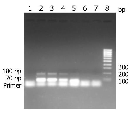
Figure 3 Detection of products of DHBV DNA positive stan-dard with qPCR by 20 g/L agarose gel electrophoresis.
Lane l: Negative control; lanes 2-7: DHBV DNA positive standard (2.76 × 10⁶-2.76 × 101 copies/µL); lane 8: DNA marker.
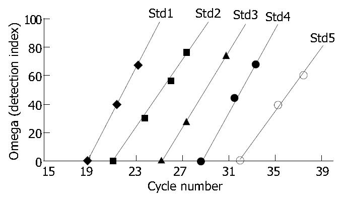
Figure 4 Amplification index standard curves of DHBV DNA positive standard.
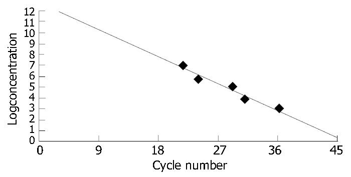
Figure 5 Concentration standard curve of DHBV DNA posi-tive standard.
- Citation: Chen YX, Huang AL, Qi ZY, Guo SH. Establishment and assessment of two methods for quantitative detection of serum duck hepatitis B virus DNA. World J Gastroenterol 2004; 10(18): 2666-2669
- URL: https://www.wjgnet.com/1007-9327/full/v10/i18/2666.htm
- DOI: https://dx.doi.org/10.3748/wjg.v10.i18.2666


