Copyright
©The Author(s) 2004.
World J Gastroenterol. Jul 15, 2004; 10(14): 2039-2044
Published online Jul 15, 2004. doi: 10.3748/wjg.v10.i14.2039
Published online Jul 15, 2004. doi: 10.3748/wjg.v10.i14.2039
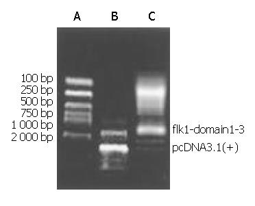
Figure 1 Clone of flk-1-domain1-3 and construction of ex-pressing vector.
A: DNA marker; B: PCR results.
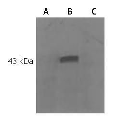
Figure 2 Functionality of the flk-1-domain 1-3 expressing vector.
A: Western blotting of protein extracted from COS7 cell; B: Western blotting of protein from COS7 transfected with vaccine; C: Western blotting of protein from COS7 transfected with pcDNA3.1 (+).
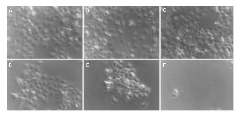
Figure 3 COS 7 cell before and after transfection with vaccine, plasmid and saline, respectively.
A, D: One day before and 2 wk after transfection with vaccine; B, E: One day before and 2 wk after transfection with plasmid; C, F: One day before and 2 wk after transfection with saline.
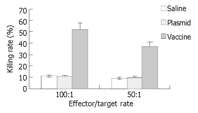
Figure 4 CTL activity of each group at different effector/target rate.
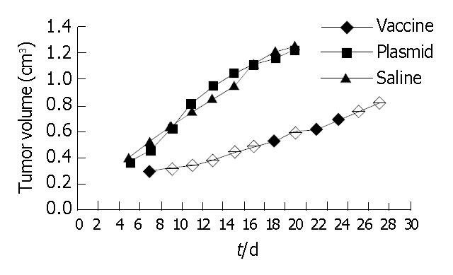
Figure 5 Curve of tumor growth in preventive group.
Figure 6 Microvessel staining by anti-CD31 antibody in preventive Group (Original magnification: × 200).
A: Vaccine subgroup; B: Plasmid control group; C: Saline control group.
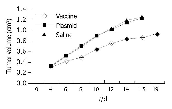
Figure 7 Curve of tumor growth in therapeutic group.
Figure 8 Microvessel staining by anti-CD31 antibody in therapeutic group.
A: Vaccine subgroup; B: Plasmid control group; C: Saline control group.
-
Citation: Lü F, Qin ZY, Yang WB, Qi YX, Li YM. A DNA vaccine against extracellular domains 1-3 of flk-1 and its immune preventive and therapeutic effects against H22 tumor cell
in vivo . World J Gastroenterol 2004; 10(14): 2039-2044 - URL: https://www.wjgnet.com/1007-9327/full/v10/i14/2039.htm
- DOI: https://dx.doi.org/10.3748/wjg.v10.i14.2039


