Copyright
©The Author(s) 2004.
World J Gastroenterol. Jun 1, 2004; 10(11): 1682-1685
Published online Jun 1, 2004. doi: 10.3748/wjg.v10.i11.1682
Published online Jun 1, 2004. doi: 10.3748/wjg.v10.i11.1682
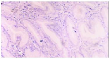
Figure 1 Expression of CDw75 in normal gastric mucosa.
No gastric mucosa cell were brown-stained either on membrane or in cytoplasm. (SP × 400).
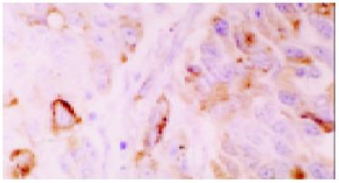
Figure 2 Weak staining of CDw75 in gastric cancer cells.
There were a few gastric cancer cells brown-stained on membrane or in cytoplasm (SP × 200).
Figure 3 Strong staining of CDw75 in gastric cancer cells.
There were many gastric cancer cells brown-stained on membrane or in cytoplasm (SP × 200).
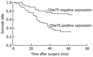
Figure 4 A Kaplan-Meier plot shows the survival rates after curative resection for gastric carcinoma patients with or with-out CDw75 expression.
- Citation: Shen L, Li HX, Luo HS, Shen ZX, Tan SY, Guo J, Sun J. CDw75 is a significant histopathological marker for gastric carcinoma. World J Gastroenterol 2004; 10(11): 1682-1685
- URL: https://www.wjgnet.com/1007-9327/full/v10/i11/1682.htm
- DOI: https://dx.doi.org/10.3748/wjg.v10.i11.1682


