Copyright
©The Author(s) 1995.
World J Gastroenterol. Oct 1, 1995; 1(1): 13-17
Published online Oct 1, 1995. doi: 10.3748/wjg.v1.i1.13
Published online Oct 1, 1995. doi: 10.3748/wjg.v1.i1.13
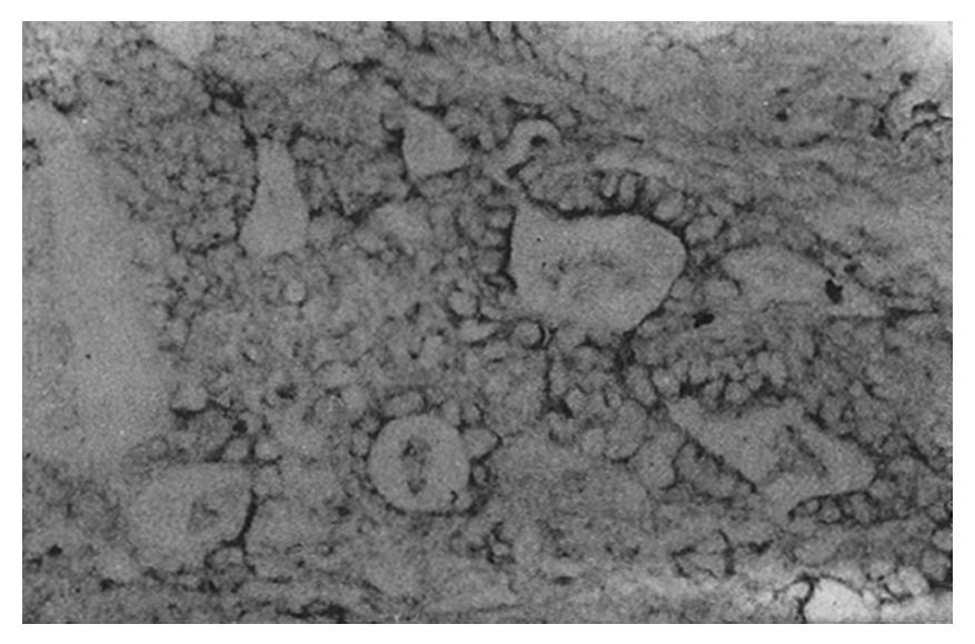
Figure 1 Tubular adenocarcinoma.
LDH-5 staining is located primarily at the luminal side of the cancer cells and partly in the cytoplasm (magnification × 200). LDH: Lactate dehydrogenase.
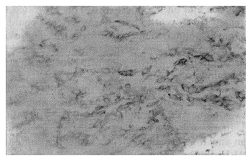
Figure 2 Poorly differentiated adenocarcinoma.
LDH-5 staining is distributed in the cytoplasm (magnification × 200). LDH: Lactate dehydrogenase.
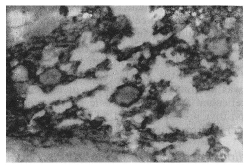
Figure 3 Tubular adenocarcinoma.
LDH-5 staining is distributed in the cytoplasmic matrix around mitochondria and the endoplasmic reticulum (magnification × 3000). LDH: Lactate dehydrogenase.
- Citation: Song YL, Yang GL, Dong YM. Immunohistochemical study of lactate dehydrogenase isoenzymes in gastric cancer. World J Gastroenterol 1995; 1(1): 13-17
- URL: https://www.wjgnet.com/1007-9327/full/v1/i1/13.htm
- DOI: https://dx.doi.org/10.3748/wjg.v1.i1.13


