Copyright
©The Author(s) 2022.
World J Clin Cases. Jun 6, 2022; 10(16): 5343-5351
Published online Jun 6, 2022. doi: 10.12998/wjcc.v10.i16.5343
Published online Jun 6, 2022. doi: 10.12998/wjcc.v10.i16.5343
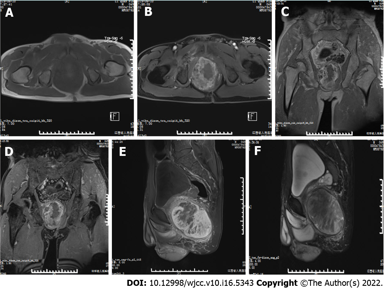
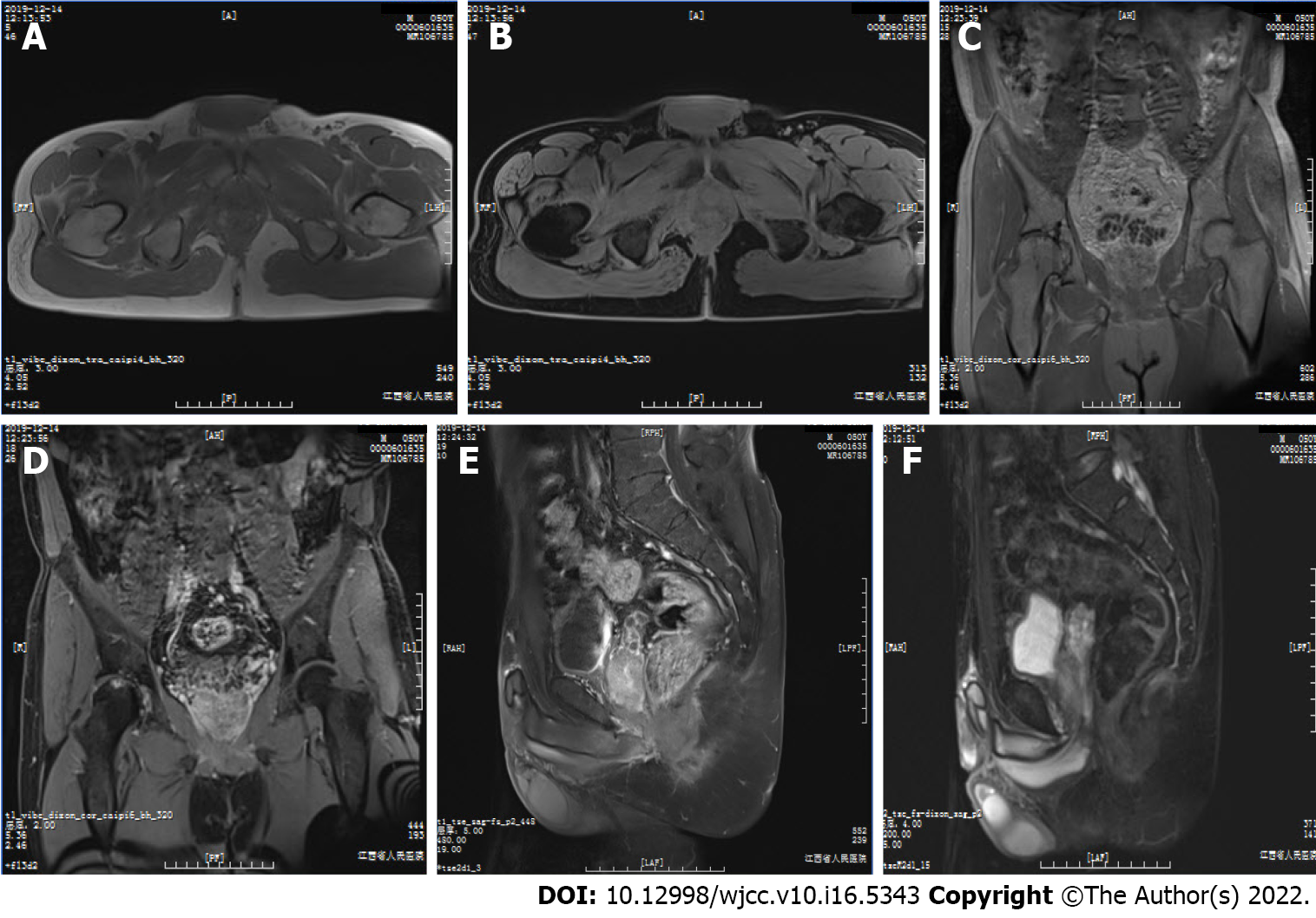
Figure 1 Magnetic resonance imaging of the mass.
A: Axial T1-weighted image (T1WI); B: Axial fat-suppressed T1 (T1FS) image; C: Coronal T1WI; D: Coronal T1FS image; E: Sagittal T1FS image; F: Sagittal T2FS image show a mass in the right pelvic cavity, which displayed predominantly isointense signals relative to the surrounding musculature on T1WI and heterogeneous signal on T1FS and T2FS. There was intralesional focal necrosis and perilesional edema.
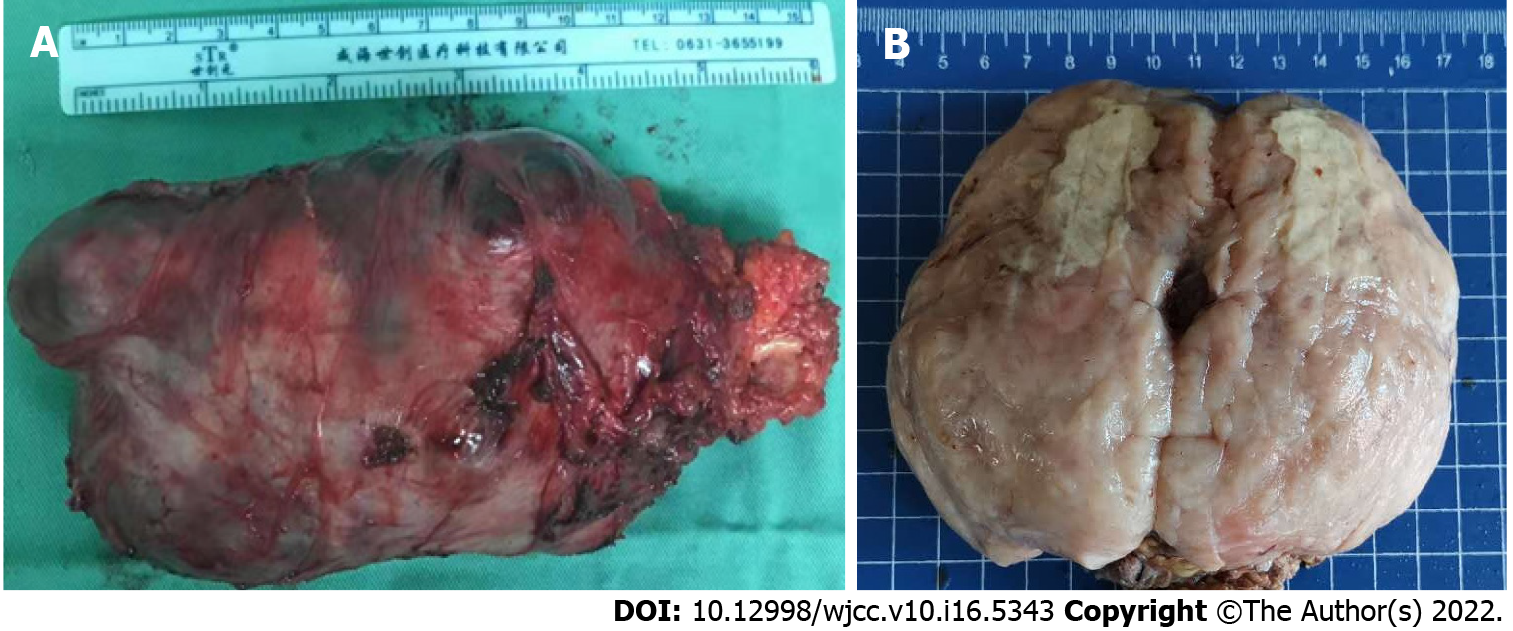
Figure 2 General observation of the mass.
A: Postoperative gross image demonstrates that the mass had an almost intact capsule with the maximum diameter of about 13 cm in size; B: General sectional view displays that the mass was solid, firm-to-elastic, and yellow-pink appearance, with focal cystic degeneration and necrosis.
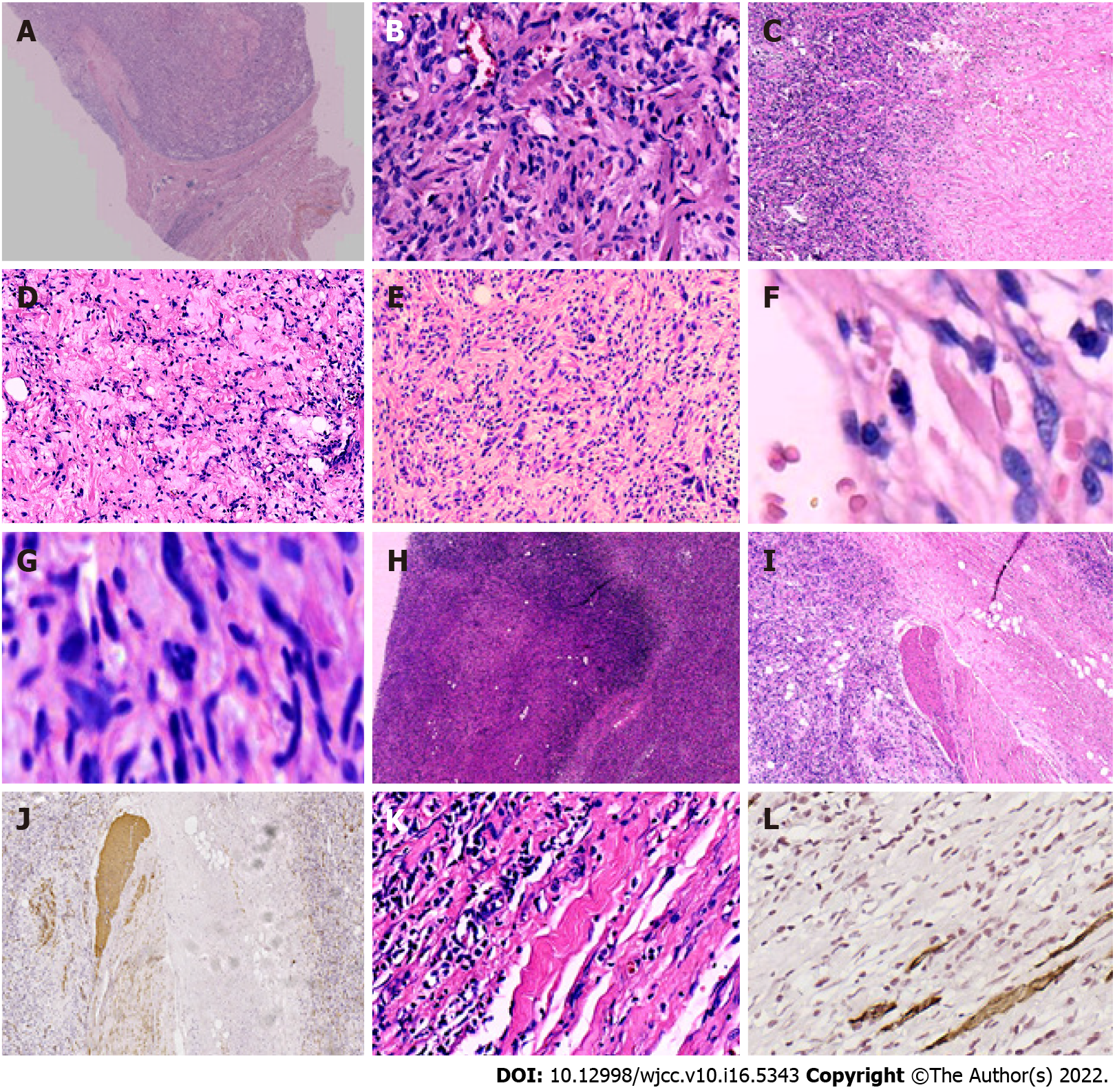
Figure 3 Histopathological features of the tumor.
A: The mass had a definite capsule; B: It was composed of short spindle to oval-shaped cells, and admixed with varying thick bundles of collagen and variable numbers of adipocytes. The cells had eosinophilic cytoplasm with indistinct cell borders and elongated nuclei with fine chromatin; C: Hyaline degeneration was visible within the tumor; D: Mucoid degeneration was visible within the tumor; E: Bizarre cells and atypic cells were unequally distributed throughout the tumor; F: Mitosis was found in the tumor cells; G: Atypical mitosis was found in the tumor cells; H: Infarction and nuclear debris were also seen inside the tumor; I and J: Smooth muscles had been infiltrated by the tumor cells; K and L: Skeletal muscles had been infiltrated by the tumor cells. Images I and K show local magnification of the black and blue boxes, respectively, in Figure A. Image J shows the infiltrated smooth muscle, which was positive for smooth muscle actin. Image L displays the invaded skeletal muscle, which was positive for desmin on immunohistochemical staining.
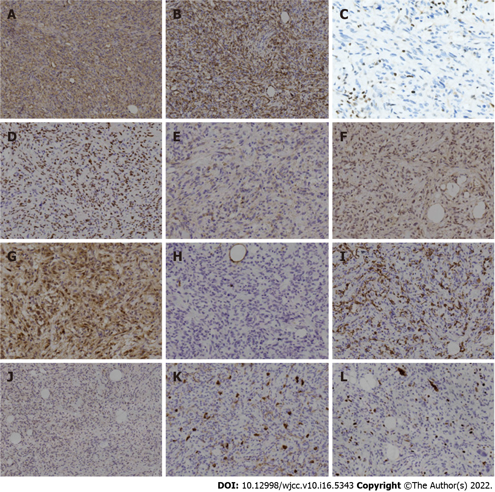
Figure 4 Immunohistochemical findings in the tumor.
A: Cells showed strongly positive staining for cluster of differentiation (CD) 34; B: Cells showed strongly positive staining for desmin; C: Lost expression of retinoblastoma 1 protein, with positive internal-control staining for lymphocytes; D: Staining was positive for estrogen receptor; E: Staining was positive for epithelial-membrane antigen; F: Staining was positive for murine double minute 2; G: Staining was positive for cyclin-dependent kinase 4; H: Staining was negative for S100; I: Staining was negative for smooth muscle actin; J: Staining was negative for signal transducer and activator of transcription 6; K: Staining was negative for CD117. Note that positive staining for CD117 appeared in the mast cells of the internal control; L: Ki-67 index was about 5%.
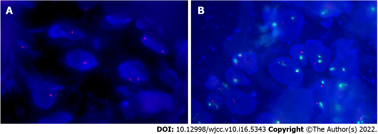
Figure 5 Molecular genetic findings in the tumor.
A: Fluorescence in situ hybridization confirmed the single and double deletion of retinoblastoma 1 alleles in neoplastic cells; B: No amplification of human homolog of murine double minute 2 in neoplastic cells.
Figure 6 Follow-up magnetic resonance imaging scan of the patient’s pelvic cavity at 6 mo post-operation.
A: T1-weighted image (T1WI); B: Axial fat-suppressed T1 (T1FS) image; C: Coronal T1WI; D: Coronal T1FS image; E: Sagittal T1FS image; F: Sagittal T2FS image showed complete removal of the mass, with normal diffusion in the patient’s pelvic cavity. There were no abnormalities except the abnormal signals from the right acetabulum and femoral head, and the ischemic necrosis of the head was considered. The size and position of pelvic visceral organs, such as the prostate, seminal vesicle, and rectum, were normal, and the bladder was well filled.
- Citation: Zeng YF, Dai YZ, Chen M. Mammary-type myofibroblastoma with infarction and atypical mitosis-a potential diagnostic pitfall: A case report. World J Clin Cases 2022; 10(16): 5343-5351
- URL: https://www.wjgnet.com/2307-8960/full/v10/i16/5343.htm
- DOI: https://dx.doi.org/10.12998/wjcc.v10.i16.5343


