Copyright
©The Author(s) 2022.
World J Clin Cases. Jun 6, 2022; 10(16): 5343-5351
Published online Jun 6, 2022. doi: 10.12998/wjcc.v10.i16.5343
Published online Jun 6, 2022. doi: 10.12998/wjcc.v10.i16.5343
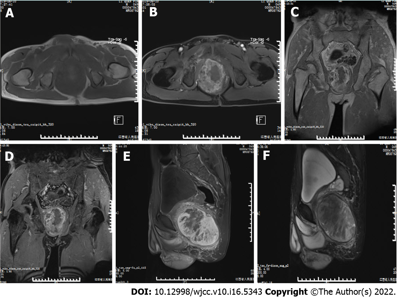
Figure 1 Magnetic resonance imaging of the mass.
A: Axial T1-weighted image (T1WI); B: Axial fat-suppressed T1 (T1FS) image; C: Coronal T1WI; D: Coronal T1FS image; E: Sagittal T1FS image; F: Sagittal T2FS image show a mass in the right pelvic cavity, which displayed predominantly isointense signals relative to the surrounding musculature on T1WI and heterogeneous signal on T1FS and T2FS. There was intralesional focal necrosis and perilesional edema.
- Citation: Zeng YF, Dai YZ, Chen M. Mammary-type myofibroblastoma with infarction and atypical mitosis-a potential diagnostic pitfall: A case report. World J Clin Cases 2022; 10(16): 5343-5351
- URL: https://www.wjgnet.com/2307-8960/full/v10/i16/5343.htm
- DOI: https://dx.doi.org/10.12998/wjcc.v10.i16.5343


