Copyright
©The Author(s) 2022.
World J Clin Cases. Jun 6, 2022; 10(16): 5343-5351
Published online Jun 6, 2022. doi: 10.12998/wjcc.v10.i16.5343
Published online Jun 6, 2022. doi: 10.12998/wjcc.v10.i16.5343
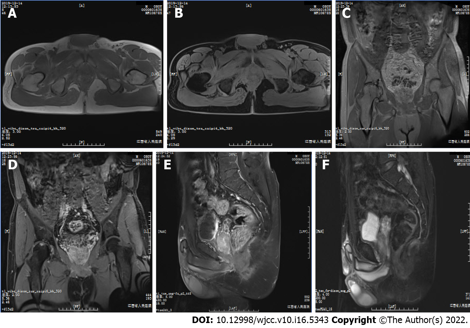
Figure 6 Follow-up magnetic resonance imaging scan of the patient’s pelvic cavity at 6 mo post-operation.
A: T1-weighted image (T1WI); B: Axial fat-suppressed T1 (T1FS) image; C: Coronal T1WI; D: Coronal T1FS image; E: Sagittal T1FS image; F: Sagittal T2FS image showed complete removal of the mass, with normal diffusion in the patient’s pelvic cavity. There were no abnormalities except the abnormal signals from the right acetabulum and femoral head, and the ischemic necrosis of the head was considered. The size and position of pelvic visceral organs, such as the prostate, seminal vesicle, and rectum, were normal, and the bladder was well filled.
- Citation: Zeng YF, Dai YZ, Chen M. Mammary-type myofibroblastoma with infarction and atypical mitosis-a potential diagnostic pitfall: A case report. World J Clin Cases 2022; 10(16): 5343-5351
- URL: https://www.wjgnet.com/2307-8960/full/v10/i16/5343.htm
- DOI: https://dx.doi.org/10.12998/wjcc.v10.i16.5343


