Copyright
©The Author(s) 2015.
World J Cardiol. Jun 26, 2015; 7(6): 299-305
Published online Jun 26, 2015. doi: 10.4330/wjc.v7.i6.299
Published online Jun 26, 2015. doi: 10.4330/wjc.v7.i6.299
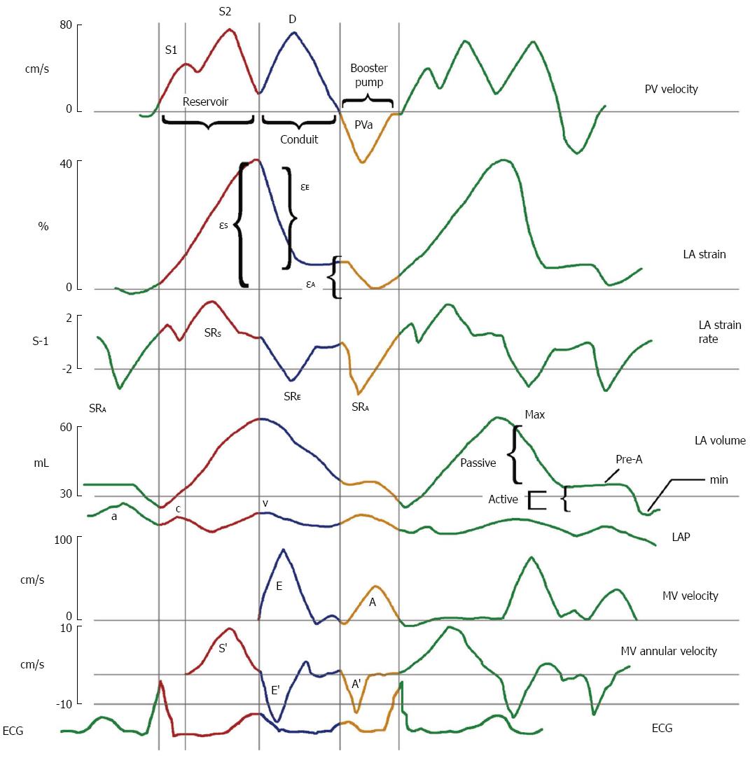
Figure 1 Left atrial physiology imaging using different methods.
The figure displays pulmonary venous (PV) velocity, left atrial (LA) strain (epsilon), LA strain rate (SR), LA volume, left atrial pressure (LAP), and mitral spectral and tissue Doppler. Displayed are two cardiac cycles and the color-coded imaging of reservoir, conduit, and booster pump functions in red, blue, and yellow lines are shown within the first cardiac cycle, respectively. Reprinted from Journal of the American College of Cardiology, Vol 63, Brian D. Hoit, Left atrial size and function: role in prognosis, 493-505, 2014 with permission from Elsevier[6].
- Citation: Kowallick JT, Lotz J, Hasenfuß G, Schuster A. Left atrial physiology and pathophysiology: Role of deformation imaging. World J Cardiol 2015; 7(6): 299-305
- URL: https://www.wjgnet.com/1949-8462/full/v7/i6/299.htm
- DOI: https://dx.doi.org/10.4330/wjc.v7.i6.299


