Copyright
©The Author(s) 2024.
World J Gastrointest Oncol. Feb 15, 2024; 16(2): 563-570
Published online Feb 15, 2024. doi: 10.4251/wjgo.v16.i2.563
Published online Feb 15, 2024. doi: 10.4251/wjgo.v16.i2.563
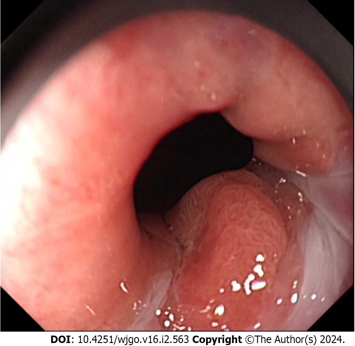
Figure 1 A representative upper endoscopic image of a tumor in the gastroesophageal junction.
An elevated mucosal lesion was found endoscopically in the distal esophageal side of the gastroesophageal junction. The metaplastic columnar epithelium extended focally in the distal esophagus above the gastroesophageal junction.
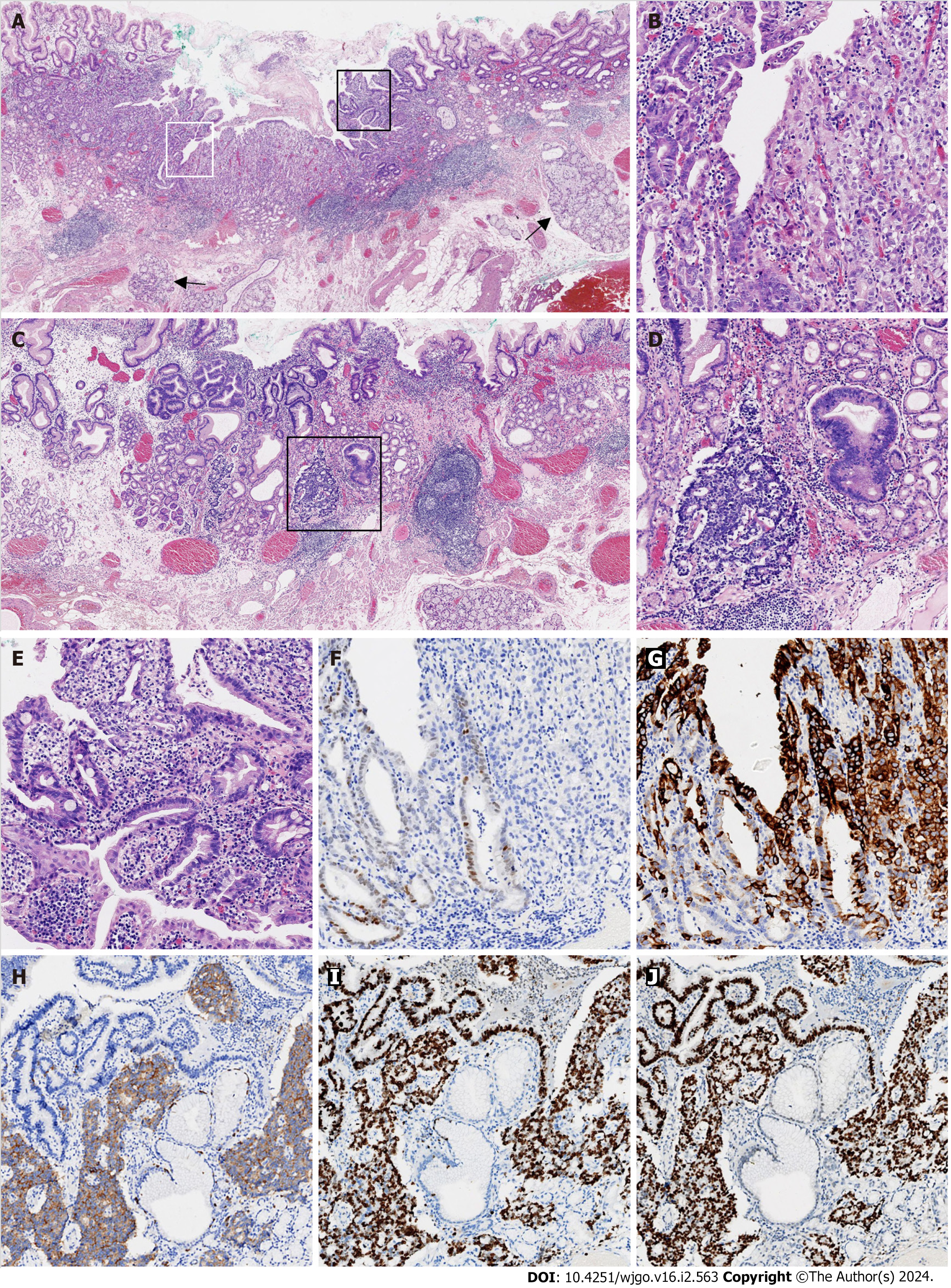
Figure 2 Histological findings of poorly differentiated adenocarcinoma mixed with a neuroendocrine carcinoma component.
A: Submucosal esophageal glands (arrow) were observed, indicating the distal esophageal location (× 20); B: The area defined by the white rectangle in Figure 2A was enlarged and showed the mixture of moderately (left) and poorly (right) differentiated adenocarcinomas (× 100); C: Adenocarcinoma with a neuroendocrine carcinoma (NEC) component arose in the cardiac mucosa (× 20); D: The area in the black rectangle in Figure 2C exhibited a mixture of adenocarcinoma (right) with NEC (left; × 100); E: The area defined by the black rectangle in Figure 2A was enlarged and demonstrated glandular dysplasia with goblet cells (× 100); F: CDX2 was weakly immunoreactive in moderately differentiated adenocarcinoma and immunonegative in poorly differentiated adenocarcinoma (× 100); G: Mucin 5AC was diffusely immunopositive in poorly differentiated adenocarcinoma and focally immunopositive in moderately differentiated adenocarcinoma (× 100); H: Synaptophysin was diffusely immunopositive in NEC, but immunonegative in adenocarcinoma (× 100); I: The Ki-67 proliferative index was approximately 90% for both the adenocarcinoma and NEC components (× 100); J: p53 was diffusely immunopositive for the adenocarcinoma and NEC components (× 100). All controls stains were adequate.
- Citation: Cheng YQ, Wang GF, Zhou XL, Lin M, Zhang XW, Huang Q. Early adenocarcinoma mixed with a neuroendocrine carcinoma component arising in the gastroesophageal junction: A case report. World J Gastrointest Oncol 2024; 16(2): 563-570
- URL: https://www.wjgnet.com/1948-5204/full/v16/i2/563.htm
- DOI: https://dx.doi.org/10.4251/wjgo.v16.i2.563


