Copyright
©The Author(s) 2018.
World J Gastrointest Endosc. Oct 16, 2018; 10(10): 283-293
Published online Oct 16, 2018. doi: 10.4253/wjge.v10.i10.283
Published online Oct 16, 2018. doi: 10.4253/wjge.v10.i10.283
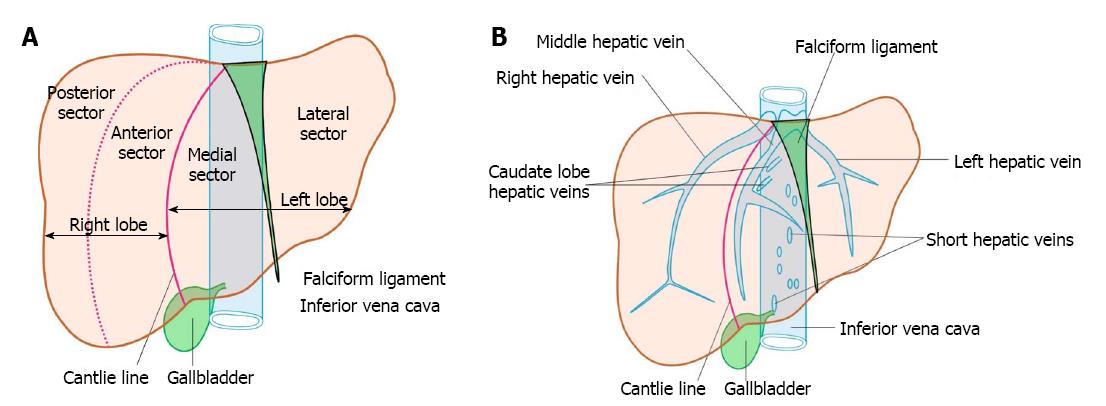
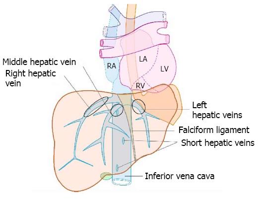
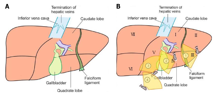
Figure 1 Anatomy of liver.
A: The four sectors of liver, i.e., right anterior, right posterior, left medial and left lateral; B: The three major veins, emerge from the posterior surface of the liver and open immediately into the supra hepatic part of inferior vena cava (IVC) just before it pierces the diaphragm. Short hepatic/accessory veins drain into lower part of IVC. The accessory veins and the caudate lobe veins join the anterior and lateral aspect of IVC.
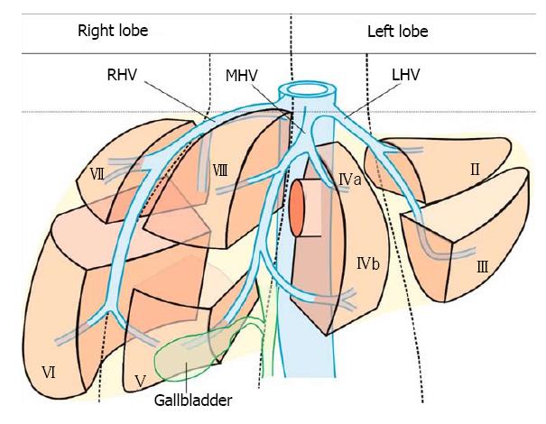
Figure 2 The segments and sectors of liver and their relationship with hepatic vein tributaries.
MHV: Middle hepatic vein; RHV: Right hepatic vein; LHV: Left hepatic vein.
Figure 3 The appearance of inferior vena cava from three stations in a rounded or elongated axis.
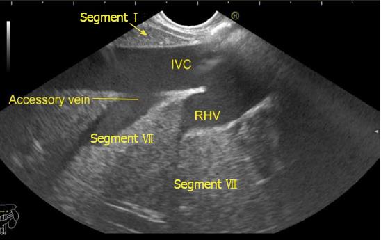
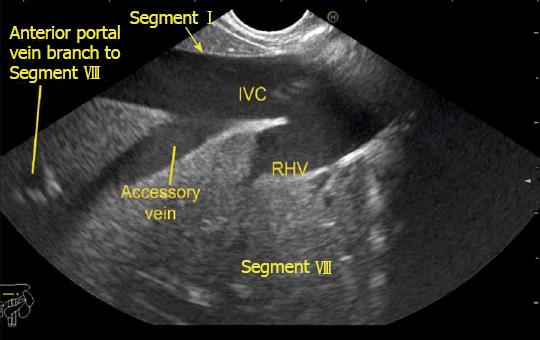
Figure 4 Inferior vena cava running parallel to the probe in a long axis from abdominal part of esophagus.
RHV: Right hepatic vein; IVC: Inferior vena cava.
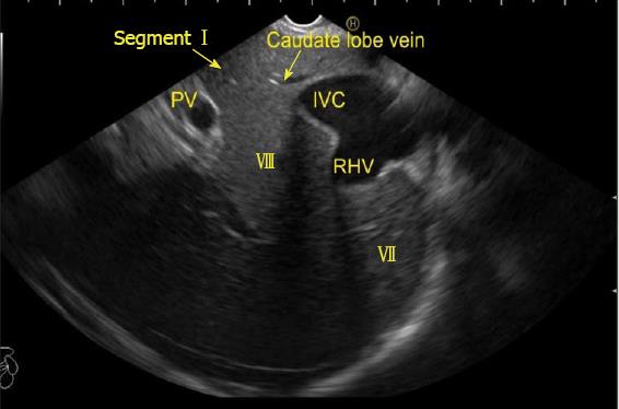
Figure 5 This figure shows inferior vena cava running parallel to the probe in a long axis.
In this image slight up angulation of the probe shows inferior vena cava in a more oval axis. PV: Portal vein; RHV: Right hepatic vein; IVC: Inferior vena cava.
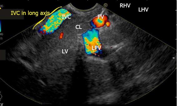
Figure 6 This figure shows inferior vena cava running from 7 o’clock position to 11 o’clock position on the far side of the screen in a long axis from the duodenal bulb.
PV: Portal vein; RHV: Right hepatic vein; LHV: Left hepatic vein; IVC: Inferior vena cava; CL: Caudate lobe; LPV: Left portal vein.
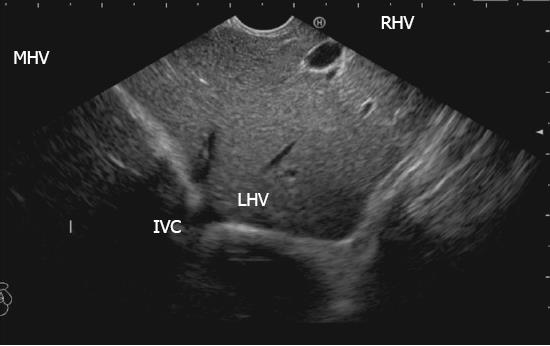
Figure 7 This figure shows inferior vena cava in a rounded axis from duodenal bulb.
MHV: Middle hepatic vein; RHV: Right hepatic vein; LHV: Left hepatic vein; IVC: Inferior vena cava.
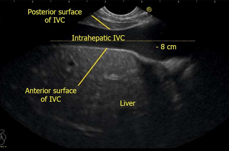
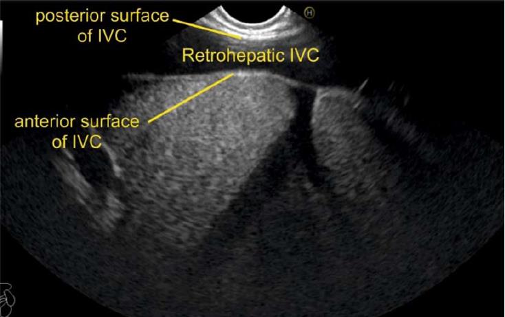
Figure 8 During imaging from the abdominal part of esophagus, the anterior surface of inferior vena cava is always found in close contact with the liver parenchyma whereas the posterior or lateral surface of the inferior vena cava is variably covered.
IVC: Inferior vena cava.
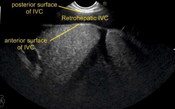
Figure 9 If the inferior vena cava is surrounded on all sides by liver parenchyma it is called as intrahepatic, if it is surrounded only anteriorly or anterolaterally it is called as retrohepatic.
In this case, right hepatic vein is seen joining the retrohepatic part of inferior vena cava. IVC: Inferior vena cava.
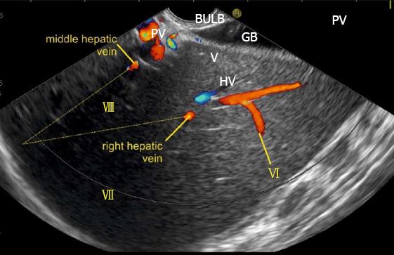
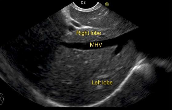
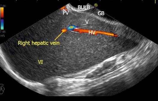
Figure 10 This image from duodenal bulb shows the right and middle hepatic vein.
The right hepatic vein goes parallel to the surface of gallbladder and the middle hepatic vein goes towards the neck of gallbladder. GB: Gall bladder.
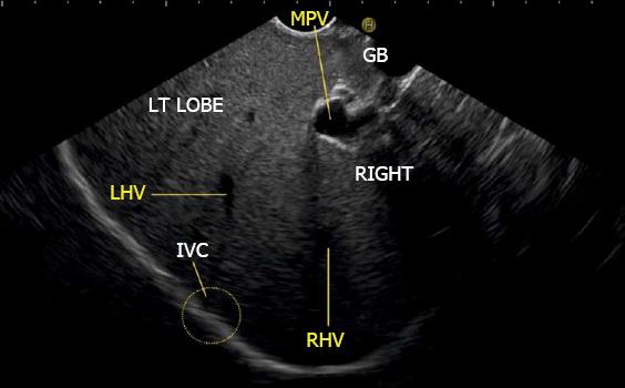
Figure 11 The inferior vena cava is not seen in this image but rotation of the scope shows the approximate area of inferior vena cava (yellow circle) where the left and right hepatic veins merge into inferior vena cava.
MHV: Middle hepatic vein; RHV: Right hepatic vein; LHV: Left hepatic vein; IVC: Inferior vena cava.
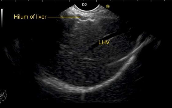
Figure 12 While imaging through the left lobe, the left hepatic vein is seen as a long vascular channel coursing towards the right side of the image into inferior vena cava which is usually seen in a rounded shape in a transverse axis.
LHV: Left hepatic vein.
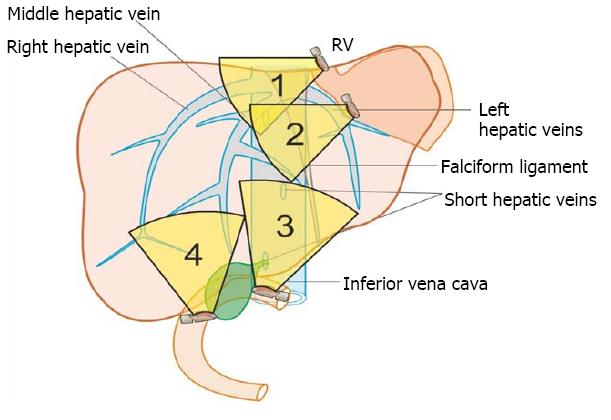
Figure 13 The imaging of hepatic veins can be done from four stations: the abdominal part of esophagus, the stomach, the duodenal bulb and the 2nd part of duodenum.
Figure 14 The images from abdominal part of esophagus shows presence of left (A), middle (B), and right hepatic vein (C) dividing the liver into four sectors.
MHV: Middle hepatic vein; RHV: Right hepatic vein; LHV: Left hepatic vein; IVC: Inferior vena cava; PV: Portal vein.
Figure 15 The tributary free part of each hepatic vein is shown.
Figure 16 The middle hepatic vein has got tributaries on the right and the left side.
The right side tributaries drain segment VIII and segment V. The left side tributaries drain segment IV. MHV: Middle hepatic vein.
Figure 17 The right hepatic vein is seen joining at an angle of around 60°.
The segment VIII is seen between hepatic vein and IVC. RHV: Right hepatic vein; IVC: Inferior vena cava.
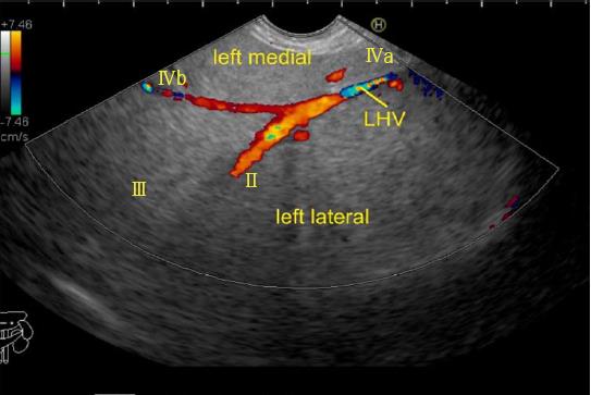
Figure 18 The left hepatic vein is seen running parallel to the probe.
The segment II, III, IVa and IVb veins are seen. In this case the imaging is done from the visceral surface of the liver and from an area close to the antrum and body. Hence, the segment IVb appears closer than segment III. LHV: Left hepatic vein.
Figure 19 Visceral surface of liver.
A: The visceral surface of the liver is shown. All the segments of liver except segment VIII are related to the visceral surface of the liver; B: The imaging from visceral surface of liver can be done from four positions.
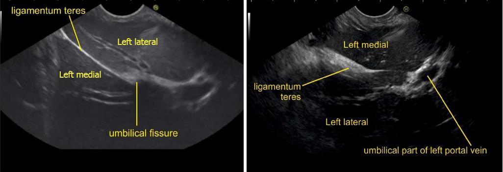
Figure 20 Imaging from visceral surface of liver shows the left lateral and left medial segment below the level of umbilical fissure separated by ligamentum teres.
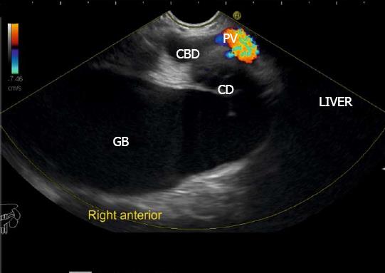
Figure 21 This image shows right anterior sector from stomach.
CBD: Common bile duct; CD: Cystic duct; GB: Gallbladder.
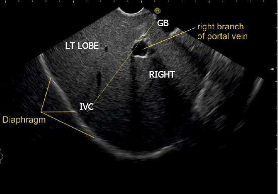
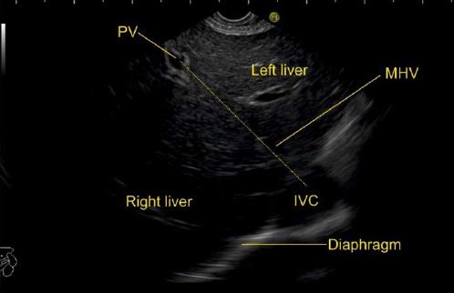
Figure 22 This image from duodenal bulb shows an imaginary line (dotted yellow line) going from inferior vena cava towards the gallbladder.
This line divides the right and left lobe of liver. The right branch of portal vein is seen as a rounded structure within the liver parenchyma in the path of this line. GB: Gallbladder; IVC: Inferior vena cava.
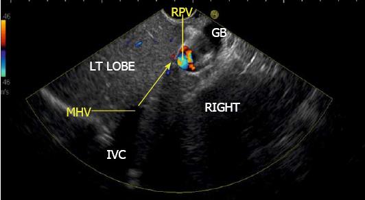
Figure 23 This image shows that the middle hepatic vein going towards the neck of gallbladder (yellow arrow).
MHV: Middle hepatic vein; GB: Gallbladder; IVC: Inferior vena cava; RPV: Right portal vein.
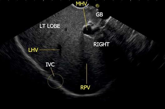
Figure 24 This image shows the course of left hepatic vein on clockwise rotation in duodenal bulb.
MHV: Middle hepatic vein; GB: Gallbladder; RHV: Right hepatic vein; LHV: Left hepatic vein; IVC: Inferior vena cava.
Figure 25 Imaging from duodenal bulb with anticlockwise rotation to visualize the right lobe.
The middle hepatic vein is seen coursing from the neck of the gallbladder and the right hepatic vein is seen coursing parallel to the upper surface of gallbladder. An imaginary line can be drawn back to the approximate position of merger into inferior vena cava which is not seen in this frame.
Figure 26 Endoscopic ultrasound from descending duodenum.
A: Figure showing right hepatic vein; B: On anticlockwise rotation from 2nd part of duodenum, the inferior vena cava gradually moves from 9 o’clock position towards 4 o’clock position. LPV: Left portal vein; PV: Portal vein; IVC: Inferior vena cava.
Figure 27 This figure shows the course of middle hepatic vein proceeding towards the portal vein by the dotted line and dividing the right anterior sector from the left medial sector.
MHV: Middle hepatic vein; PV: Portal vein; IVC: Inferior vena cava.
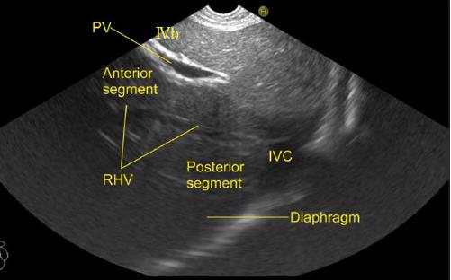
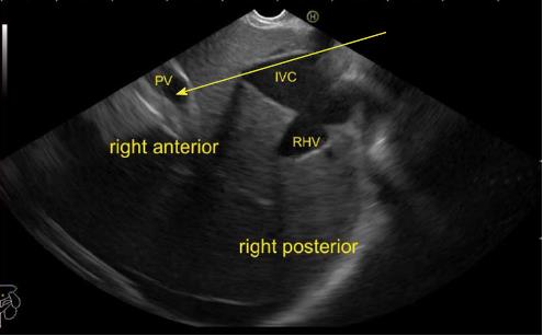
Figure 28 This figure shows the course of right hepatic vein dividing the anterior and posterior sector of right lobe of liver.
PV: Portal vein; RHV: Right hepatic vein; IVC: Inferior vena cava.
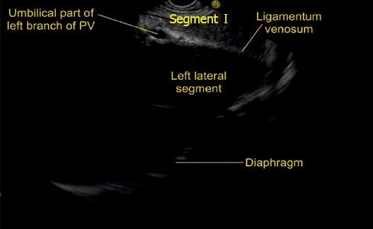
Figure 29 This figure shows the course of ligamentum venosum proceeding towards the umbilical part of portal vein and dividing the left lateral segment from the caudate lobe.
The separation of left lateral and left medial segment is done by the course of left hepatic vein but more posteriorly near the liver hilum the ligamentum venosum separates left lateral segment from the caudate lobe. PV: Portal vein.
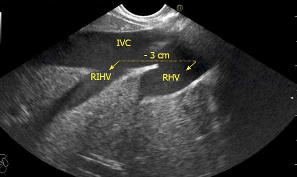
Figure 30 This image shows the presence of prominent right inferior hepatic vein below the right main hepatic vein.
This vein has a size almost similar to main right hepatic vein and provides segmental drainage of a complete segment. The distance between right hepatic vein from the right inferior hepatic vein in this image is 3 cm. IVC: Inferior vena cava; RHV: Right hepatic vein; RIHV: Right inferior hepatic vein.
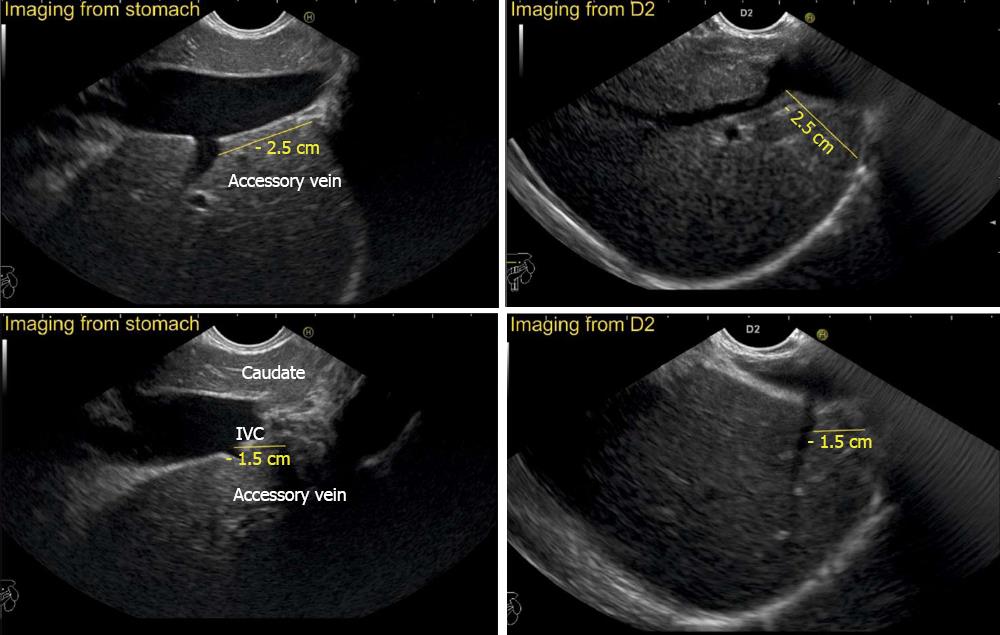
Figure 31 This image shows the right inferior accessory hepatic vein draining the right posterior sector.
The presence of liver parenchyma above the joining of the vein points to retro rather than suprahepatic course of the vein. In this case the main right hepatic vein was absent and all the venous drainage was provided by the accessory vein. IVC: Inferior vena cava.
Figure 32 In this case, two accessory veins are seen about 1.
5 cm and 2.5 cm below the diaphragm. A comparative imaging of the same accessory vein is shown from stomach and second part of duodenum.
Figure 33 The imaging is done from abdominal part of esophagus and the main portal vein is seen on the far side of the screen.
A possible communication is shown between the inferior vena cava and the main portal vein by the arrow. PV: Portal vein; RHV: Right hepatic vein; IVC: Inferior vena cava.
- Citation: Sharma M, Somani P, Rameshbabu CS. Linear endoscopic ultrasound evaluation of hepatic veins. World J Gastrointest Endosc 2018; 10(10): 283-293
- URL: https://www.wjgnet.com/1948-5190/full/v10/i10/283.htm
- DOI: https://dx.doi.org/10.4253/wjge.v10.i10.283


