Copyright
©The Author(s) 2018.
World J Gastrointest Endosc. Oct 16, 2018; 10(10): 283-293
Published online Oct 16, 2018. doi: 10.4253/wjge.v10.i10.283
Published online Oct 16, 2018. doi: 10.4253/wjge.v10.i10.283
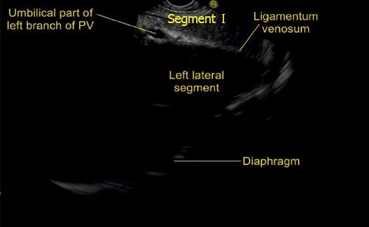
Figure 29 This figure shows the course of ligamentum venosum proceeding towards the umbilical part of portal vein and dividing the left lateral segment from the caudate lobe.
The separation of left lateral and left medial segment is done by the course of left hepatic vein but more posteriorly near the liver hilum the ligamentum venosum separates left lateral segment from the caudate lobe. PV: Portal vein.
- Citation: Sharma M, Somani P, Rameshbabu CS. Linear endoscopic ultrasound evaluation of hepatic veins. World J Gastrointest Endosc 2018; 10(10): 283-293
- URL: https://www.wjgnet.com/1948-5190/full/v10/i10/283.htm
- DOI: https://dx.doi.org/10.4253/wjge.v10.i10.283


