Copyright
©The Author(s) 2018.
World J Gastrointest Endosc. Oct 16, 2018; 10(10): 283-293
Published online Oct 16, 2018. doi: 10.4253/wjge.v10.i10.283
Published online Oct 16, 2018. doi: 10.4253/wjge.v10.i10.283
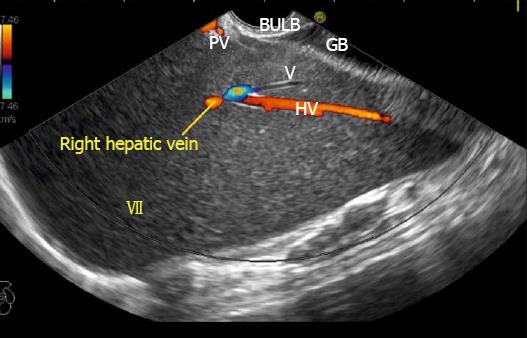
Figure 25 Imaging from duodenal bulb with anticlockwise rotation to visualize the right lobe.
The middle hepatic vein is seen coursing from the neck of the gallbladder and the right hepatic vein is seen coursing parallel to the upper surface of gallbladder. An imaginary line can be drawn back to the approximate position of merger into inferior vena cava which is not seen in this frame.
- Citation: Sharma M, Somani P, Rameshbabu CS. Linear endoscopic ultrasound evaluation of hepatic veins. World J Gastrointest Endosc 2018; 10(10): 283-293
- URL: https://www.wjgnet.com/1948-5190/full/v10/i10/283.htm
- DOI: https://dx.doi.org/10.4253/wjge.v10.i10.283


