Copyright
©The Author(s) 2018.
World J Gastrointest Endosc. Oct 16, 2018; 10(10): 283-293
Published online Oct 16, 2018. doi: 10.4253/wjge.v10.i10.283
Published online Oct 16, 2018. doi: 10.4253/wjge.v10.i10.283
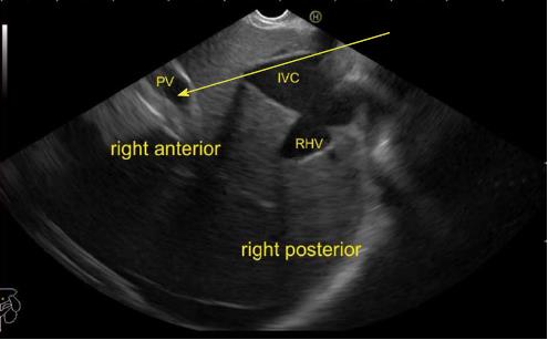
Figure 33 The imaging is done from abdominal part of esophagus and the main portal vein is seen on the far side of the screen.
A possible communication is shown between the inferior vena cava and the main portal vein by the arrow. PV: Portal vein; RHV: Right hepatic vein; IVC: Inferior vena cava.
- Citation: Sharma M, Somani P, Rameshbabu CS. Linear endoscopic ultrasound evaluation of hepatic veins. World J Gastrointest Endosc 2018; 10(10): 283-293
- URL: https://www.wjgnet.com/1948-5190/full/v10/i10/283.htm
- DOI: https://dx.doi.org/10.4253/wjge.v10.i10.283


