Copyright
©2012 Baishideng Publishing Group Co.
World J Hepatol. Dec 27, 2012; 4(12): 356-364
Published online Dec 27, 2012. doi: 10.4254/wjh.v4.i12.356
Published online Dec 27, 2012. doi: 10.4254/wjh.v4.i12.356
Figure 1 Genes upregulated in activation of hepatic stellate cells.
A: Rat hepatic stellate cells (HSCs) were isolated and cultured for 2 d (quiescent HSCs) or for 7 d (activated HSCs) and total RNA was analyzed for gene expression by microarrays in two independent experiments. The fold increase in transcript level and P values for the selected genes is shown; B: Expression of cytokine receptor-like factor 1 (CRLF1), cardiotrophin-like cytokine factor 1 (CLCF1) and ciliary neurotrophic factor receptor (CNTFR) in quiescent and activated HSCs was estimated by reverse transcription-polymerase chain reaction. Q vs A: Quiescent HSCs vs activated HSCs; IL-6: Interleukin-6; IL6ST: Interleukin 6 signal transducer; IL6R: Interleukin 6 receptor; CNTF: Ciliary neurotrophic factor.
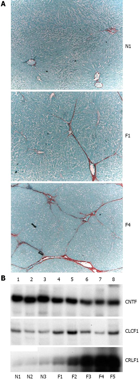
Figure 2 Expression of cytokine receptor-like factor 1 in fibrotic livers.
A: Sirius red staining of liver slices from three representative animals; N1: Normal liver; F1: Liver with moderate degree of fibrosis induced by CCl4; F4: Liver with high degree of fibrosis induced by CCl4; B: Reverse transcription-polymerase chain reaction analysis of expression of cytokine receptor-like factor 1 (CRLF1), cardiotrophin-like cytokine factor 1 (CLCF1) in three normal livers (N1-N3) and five fibrotic livers (F1-F5).
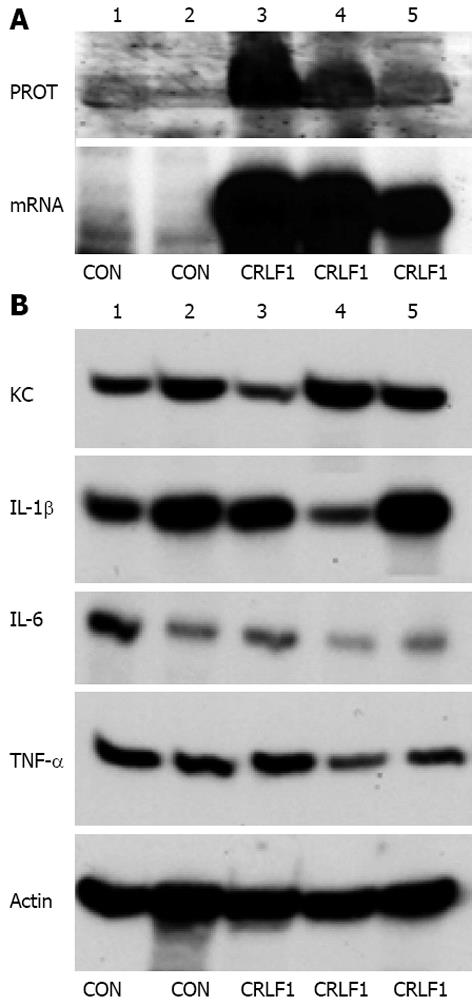
Figure 3 Overexpression of cytokine receptor-like factor 1 in normal liver does not increase expression of profibrotic cytokines.
A: Overexpression of cytokine receptor-like factor 1 (CRLF1). Control adenovirus was injected into tail vein of two mice and adenovirus expressing cardiotrophin-like cytokine factor 1 (CLCF1) gene was injected into three mice. After three days liver proteins and RNA were extracted and expression of CLRF1 protein was analyzed by Western blot (top panel) and CLRF1 mRNA was analyzed by reverse transcription-polymerase chain reaction (RT-PCR) (bottom panel); B: Expression of profibrotic cytokines. Expression of the indicated profibrotic cytokines was analyzed by RT-PCR in the control (CON) and CRLF1 overexpressing livers. PROT: Protein; CNTFR: Ciliary neurotrophic factor receptor; IL: Interleukin; TNF-α: Tumour necrosis factor-α; KC: Chemokine.
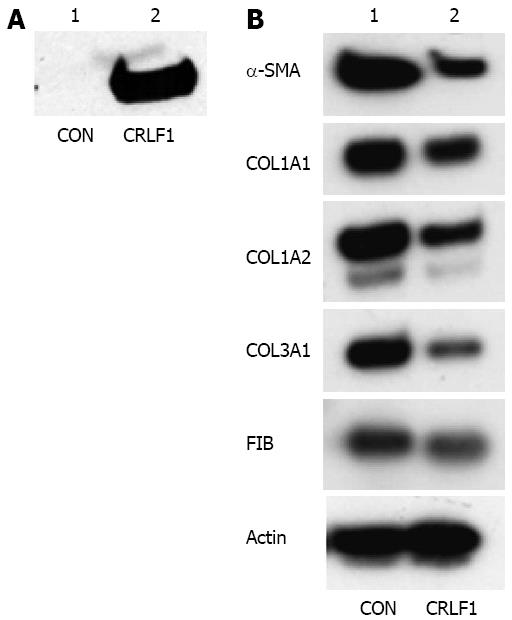
Figure 4 Cytokine receptor-like factor 1 decreases expression of type III collagen by hepatic stellate cells.
A. Expression of adenovirus delivered cytokine receptor-like factor 1 (CRLF1) in the cellular medium. HEK293 cells were transduced with control or CRLF1 expressing adenovirus. One day after transduction, CRLF1 protein was measured in the cellular medium by Western blotting; B: Stimulation of hepatic stellate cells (HSCs) with CRLF1. HSCs at day 2 after isolation were transduced with control or CRLF1 adenovirus. At day 7, expression of α-smooth muscle actin (α-SMA), collagen α1 (I), collagen α2 (I), collagen α1 (III) and fibronectin (FIB) mRNAs was analyzed by reverse transcription-polymerase chain reaction. CON: Control; COL1A1: Collagen, type I, α1; COL1A2: Collagen, type I, α2; COL3A1: Collagen, type III, α1.
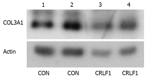
Figure 5 Cytokine receptor-like factor 1 decreases type III collagen expression in the whole liver.
RNA was extracted from two control livers and two livers that overexpressed cytokine receptor-like factor 1 (CRLF1) after adenoviral delivery and expression of collagen α1 (III) mRNA was analyzed by reverse transcription-polymerase chain reaction. CON: Control; COL3A1: Collagen, type III, α1.
- Citation: Stefanovic L, Stefanovic B. Role of cytokine receptor-like factor 1 in hepatic stellate cells and fibrosis. World J Hepatol 2012; 4(12): 356-364
- URL: https://www.wjgnet.com/1948-5182/full/v4/i12/356.htm
- DOI: https://dx.doi.org/10.4254/wjh.v4.i12.356


