Copyright
©The Author(s) 2016.
World J Stem Cells. Oct 26, 2016; 8(10): 342-354
Published online Oct 26, 2016. doi: 10.4252/wjsc.v8.i10.342
Published online Oct 26, 2016. doi: 10.4252/wjsc.v8.i10.342
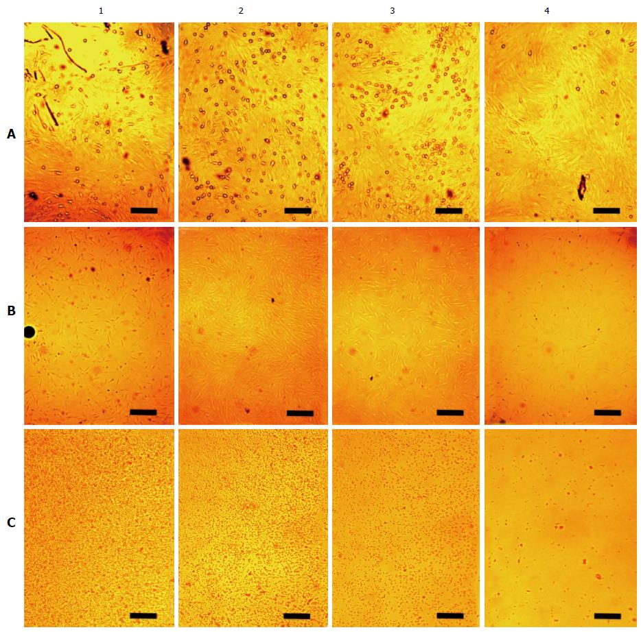
Figure 5 Arrangements of living unstained cells in affecting the magnetized needle.
Snapshots were carried out in four perpendicular directions from the center of the cultural dish in four concentric areas fixed width (Figure 2). In columns: The central region (1); the area near the central region (2); the area near the fringe region (3); the fringe region (4). In rows: A: Arrangements at the dish bottom of rBMSCs cultured for 6 d under the influence of the magnetized needle (the needle in the center of the culture dish). The small rounded cells of approximately a micron are seen over the cell monolayer. They are unevenly distributed, depending on their distance from the center of the dish. The bar is 10 μm; B: Arrangements of hBMSCs cultured for 1 d under the influence of the needle (the needle in the center of the culture dish). The dark round trace of the needle is seen on the left edge of the photo B1. The bar is 25 μm; C: Migration of human mononuclear leukocytes during a day without expo-sure to the needle. The bar is 10 μm. rBMSCs: Rat bone marrow-derived stromal stem cells; hBMSCs: Human bone marrow-derived stromal stem cells.
- Citation: Emelyanov AN, Borisova MV, Kiryanova VV. Model acupuncture point: Bone marrow-derived stromal stem cells are moved by a weak electromagnetic field. World J Stem Cells 2016; 8(10): 342-354
- URL: https://www.wjgnet.com/1948-0210/full/v8/i10/342.htm
- DOI: https://dx.doi.org/10.4252/wjsc.v8.i10.342


