
















©The Author(s) 2015.
World J Stem Cells. Sep 26, 2015; 7(8): 1127-1136
Published online Sep 26, 2015. doi: 10.4252/wjsc.v7.i8.1127
Published online Sep 26, 2015. doi: 10.4252/wjsc.v7.i8.1127
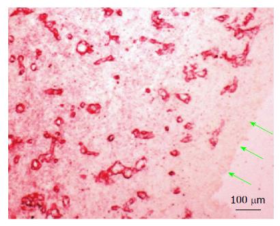
Figure 5 Vascularity of the medial layer of the subcutaneous tissue patch 14 d after implantation of the inert body.
Microvessels were revealed by immune-staining for collagen type IV, shown earlier to stain newly formed blood vessels[15]. Green arrows point to the inside aspect of the tissue patch in contact with the inert body. Note the absence of blood vessels in the inner layer.
- Citation: Garcia-Gomez I, Gudehithlu KP, Arruda JAL, Singh AK. Autologous tissue patch rich in stem cells created in the subcutaneous tissue. World J Stem Cells 2015; 7(8): 1127-1136
- URL: https://www.wjgnet.com/1948-0210/full/v7/i8/1127.htm
- DOI: https://dx.doi.org/10.4252/wjsc.v7.i8.1127