Copyright
©The Author(s) 2003.
World J Gastroenterol. May 15, 2003; 9(5): 946-950
Published online May 15, 2003. doi: 10.3748/wjg.v9.i5.946
Published online May 15, 2003. doi: 10.3748/wjg.v9.i5.946
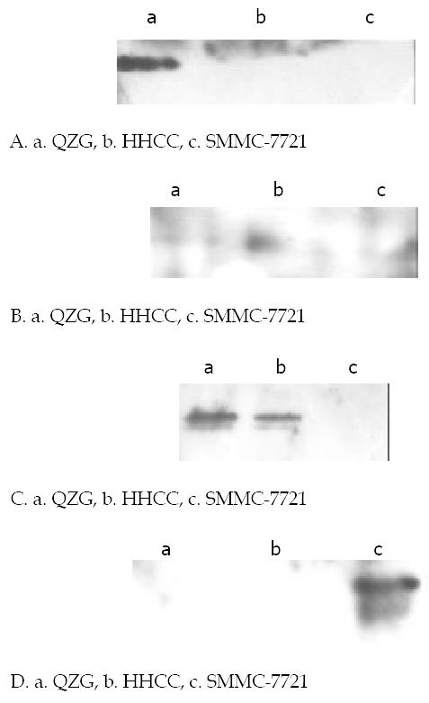
Figure 4 Western blot analysis of phosphorylation on tyrosine of CX32, CX43 proteins in various cells lines.
Cells were harvested and lysed, equal amount of cell lystates were resolved of SDS-PAGE, transferred to NC membranes, and then probed with anti-phophorylation tyrosine mAb 4G10. (A). Expressions of CX32 proteins in cell lines with special anti-CX32 mAb. CX32 showed high immunoblot signal only in normal hepatocyte cell line QZG with very slight signal in hepatocarcinoma cell lines HHCC, SMMC-7721. (B). Expressions of CX43 proteins in cell lines with special anti-CX43 mAb. CX43 apperared in both QZG and SMMC-7721 cells but no in HHCC. (C). Phosphorylation on tyrosine of CX43 with special anti-phosphrylation tyrosine 4G10, unphosphorylation appeared in QZG cells even they showed high level expression of CX32, CX43. (D). Phosphorylated tyrosine of CX43 protein was detected in SMMC-7721 cells.
- Citation: Ma XD, Ma X, Sui YF, Wang WL, Wang CM. Signal transduction of gap junctional genes, connexin32, connexin43 in human hepatocarcinogenesis. World J Gastroenterol 2003; 9(5): 946-950
- URL: https://www.wjgnet.com/1007-9327/full/v9/i5/946.htm
- DOI: https://dx.doi.org/10.3748/wjg.v9.i5.946


