Copyright
©The Author(s) 2003.
World J Gastroenterol. Oct 15, 2003; 9(10): 2226-2231
Published online Oct 15, 2003. doi: 10.3748/wjg.v9.i10.2226
Published online Oct 15, 2003. doi: 10.3748/wjg.v9.i10.2226
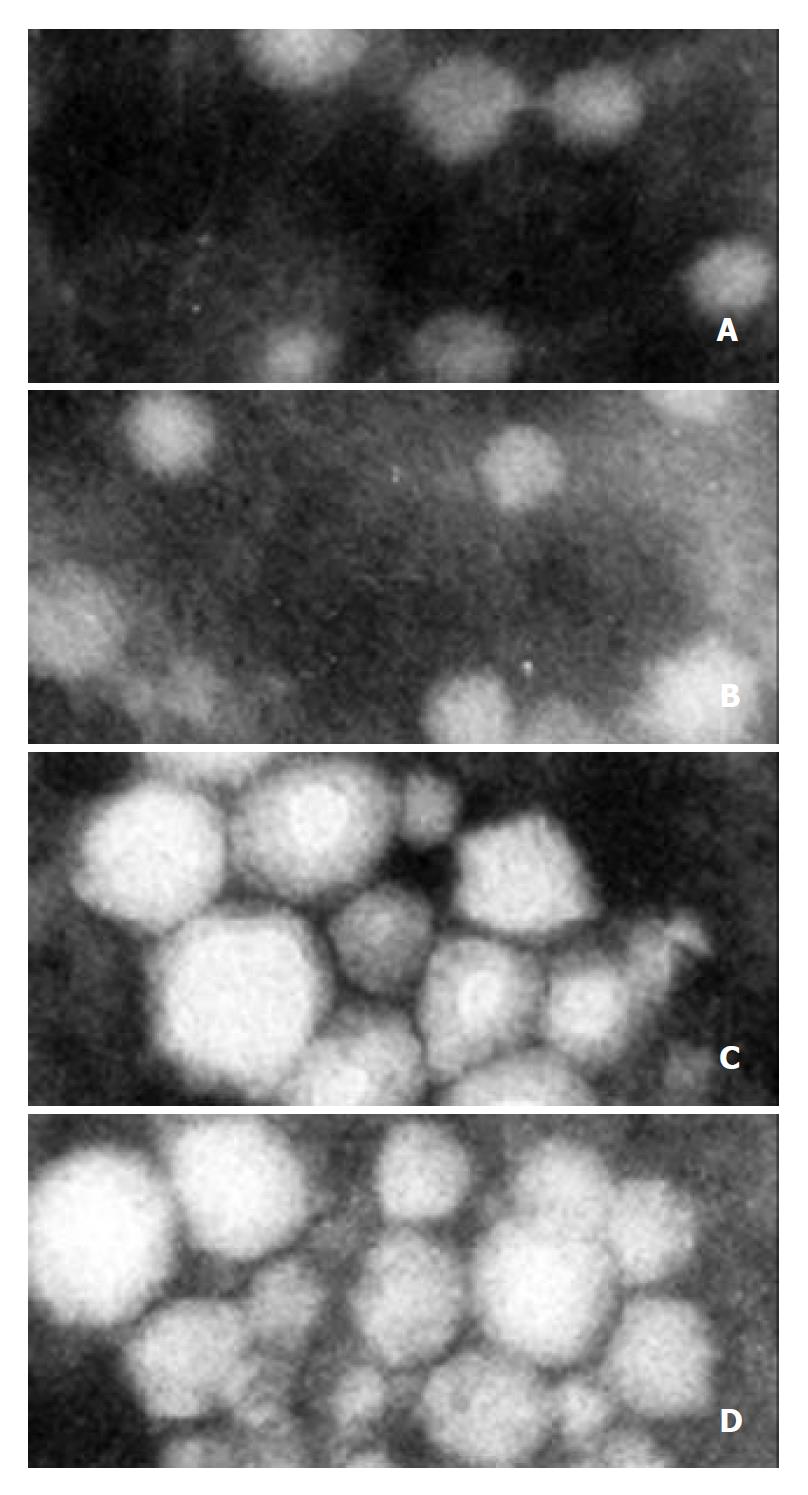
Figure 11 Electron microscopy and immune aggregation elec-tron microscopy analysis of VLPs (negative staining).
A and B (× 100000): VLPs isolated from Sf9 cells infected with reBV/CS and reBV/CE1E2, respectively. C and D (× 150000): Immune aggregation of VLPs from A and B with anti-HCV serum, respectively.
- Citation: Zhao W, Liao GY, Jiang YJ, Jiang SD. No requirement of HCV 5’NCR for HCV-like particles assembly in insect cells. World J Gastroenterol 2003; 9(10): 2226-2231
- URL: https://www.wjgnet.com/1007-9327/full/v9/i10/2226.htm
- DOI: https://dx.doi.org/10.3748/wjg.v9.i10.2226


