Copyright
©The Author(s) 2001.
World J Gastroenterol. Feb 15, 2001; 7(1): 74-79
Published online Feb 15, 2001. doi: 10.3748/wjg.v7.i1.74
Published online Feb 15, 2001. doi: 10.3748/wjg.v7.i1.74
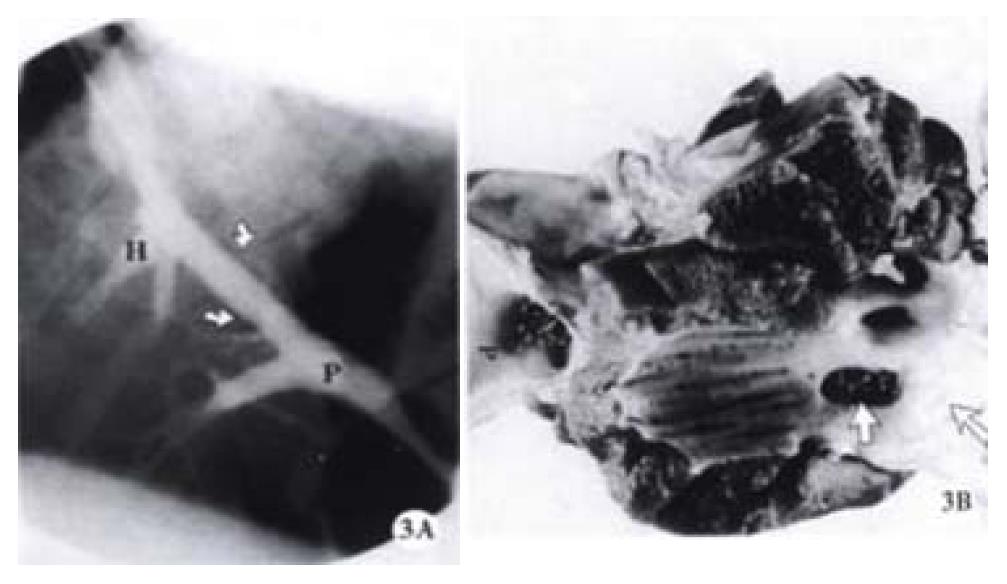
Figure 3 a.
An occluded TIPS shunt from a pig using a Cordis stent. The immediate portal venography after TIPS shows a patent TIPS shunt (arrows). b. The occluded shunt was confirmed by the portography obtained immediately before the euthanasia in two weeks after the stent placement. The gross examination of the specimen demonstrates that the hepatic end of the stent wedges into the liver parenchyma and the proximal end of the shunt is totally occluded by the healed hepatic vein (arrows), while the portal end (P) of the stent keeps patent.
- Citation: Teng GJ, Bettmann MA, Hoopes PJ, Yang L. Comparison of a new stent and Wallstent for transjugular intrahepatic portosystemic shunt in a porcine model. World J Gastroenterol 2001; 7(1): 74-79
- URL: https://www.wjgnet.com/1007-9327/full/v7/i1/74.htm
- DOI: https://dx.doi.org/10.3748/wjg.v7.i1.74


