Copyright
©The Author(s) 1998.
World J Gastroenterol. Jun 15, 1998; 4(3): 206-209
Published online Jun 15, 1998. doi: 10.3748/wjg.v4.i3.206
Published online Jun 15, 1998. doi: 10.3748/wjg.v4.i3.206
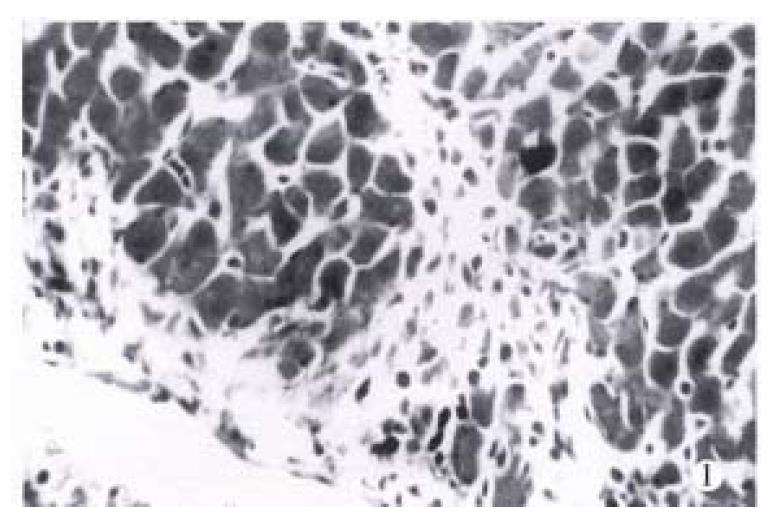
Figure 1 At the end of the 3rd week, the structure of liver lobules was defined well, the portal tracts were enlarged mildly with increased ECM and proliferated mesenchymal cells.
The infiltration of eosinophils was visible in these areas. HE × 200
- Citation: Huang ZG, Zhai WR, Zhang YE, Zhang XR. Study of heteroserum-induced rat liver fibrosis model and its mechanism. World J Gastroenterol 1998; 4(3): 206-209
- URL: https://www.wjgnet.com/1007-9327/full/v4/i3/206.htm
- DOI: https://dx.doi.org/10.3748/wjg.v4.i3.206


