Copyright
©The Author(s) 1998.
World J Gastroenterol. Apr 15, 1998; 4(2): 158-161
Published online Apr 15, 1998. doi: 10.3748/wjg.v4.i2.158
Published online Apr 15, 1998. doi: 10.3748/wjg.v4.i2.158
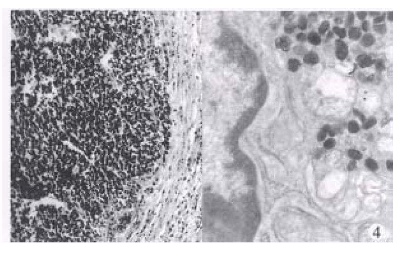
Figure 4 Type 3 neuroendocrine carcinoma.
Tumor cells are small and round, mitoses are abundant. Focal necrosis was found. HE × 250 (left). Ultrastructural appearance of the neuroendocrine carcinoma with characteristic secrectory granules. × 28000 (right)
- Citation: Yu JY, Wang LP, Meng YH, Hu M, Wang JL, Bordi C. Classification of gastric neuroendocrine tumors and its clinicopathologic significance. World J Gastroenterol 1998; 4(2): 158-161
- URL: https://www.wjgnet.com/1007-9327/full/v4/i2/158.htm
- DOI: https://dx.doi.org/10.3748/wjg.v4.i2.158


