Copyright
©The Author(s) 2025.
World J Gastroenterol. May 21, 2025; 31(19): 104897
Published online May 21, 2025. doi: 10.3748/wjg.v31.i19.104897
Published online May 21, 2025. doi: 10.3748/wjg.v31.i19.104897
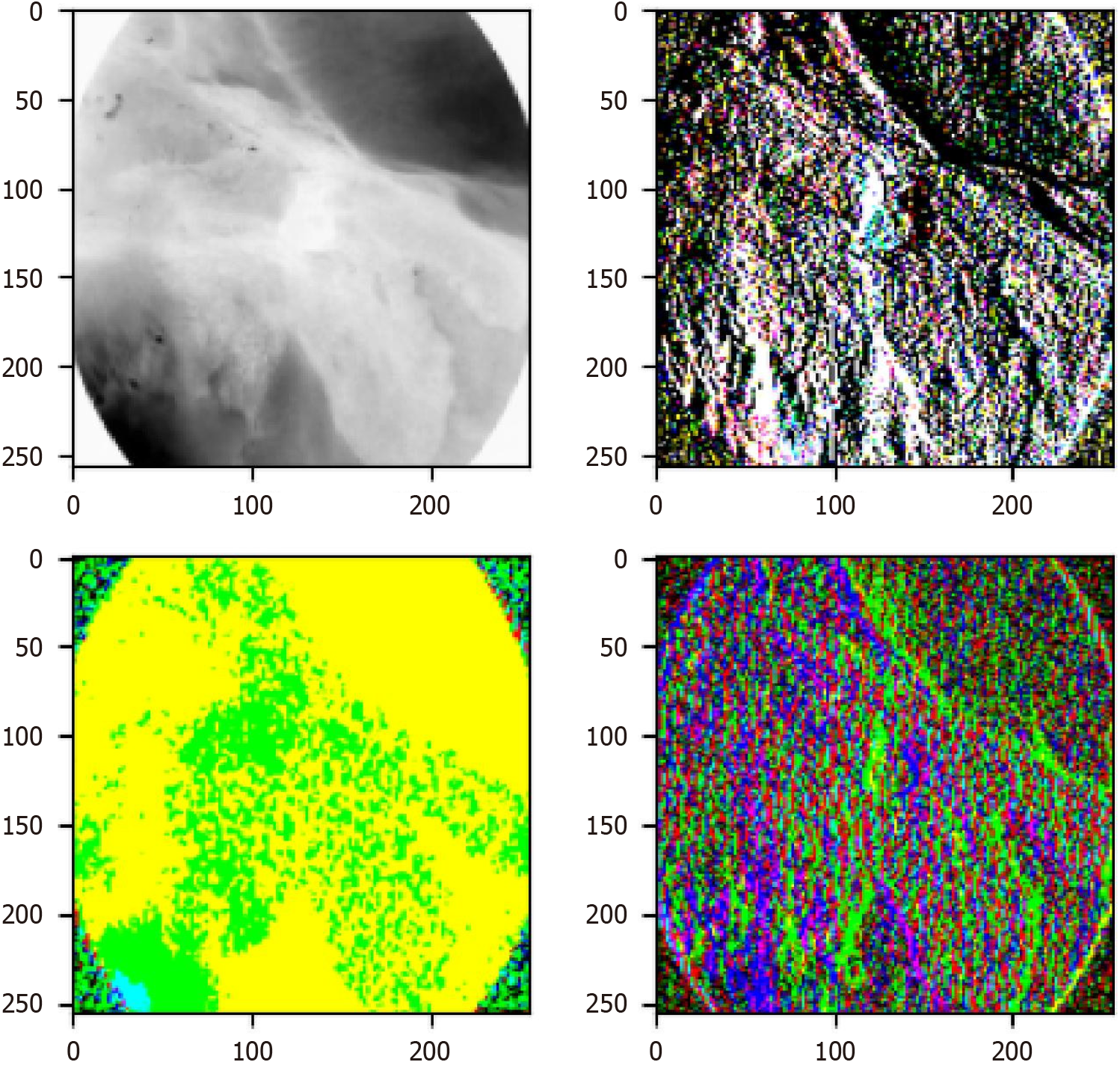
Figure 8 Visualization of wavelet block extracting different frequency domain features.
The top left image represents the low-frequency sub-band, while the other images depict high-frequency sub-bands. After processing the different sub-band images, they can be combined into a single image through the inverse wavelet transform, allowing for the extraction of additional information. After processing different sub-bands and applying the inverse wavelet transform, these components can be recombined into a single image, thereby extracting richer information. This demonstrates how wavelet transform decomposes an original image into low- and high-frequency sub-bands, where the low-frequency sub-band preserves the primary structure and global information, while the high-frequency sub-bands capture fine details and edge features. By processing and reconstructing these sub-bands, the multi-scale feature representation of the image is enhanced without losing critical information. This process significantly improves the model’s ability to detect subtle pathological changes, which is crucial for esophageal cancer diagnosis. Particularly in early-stage esophageal cancer, small and hidden morphological differences among subtypes can be better captured. Therefore, the figure not only validates the effectiveness of wavelet transform in feature extraction but also provides strong technical support for the study. The findings demonstrate that the Wave-Vision Transformer model, by integrating multi-scale feature fusion, substantially enhances the accuracy and robustness of esophageal cancer diagnosis[23].
- Citation: Wei W, Zhang XL, Wang HZ, Wang LL, Wen JL, Han X, Liu Q. Application of deep learning models in the pathological classification and staging of esophageal cancer: A focus on Wave-Vision Transformer. World J Gastroenterol 2025; 31(19): 104897
- URL: https://www.wjgnet.com/1007-9327/full/v31/i19/104897.htm
- DOI: https://dx.doi.org/10.3748/wjg.v31.i19.104897


