Copyright
©The Author(s) 2025.
World J Gastroenterol. May 21, 2025; 31(19): 104897
Published online May 21, 2025. doi: 10.3748/wjg.v31.i19.104897
Published online May 21, 2025. doi: 10.3748/wjg.v31.i19.104897
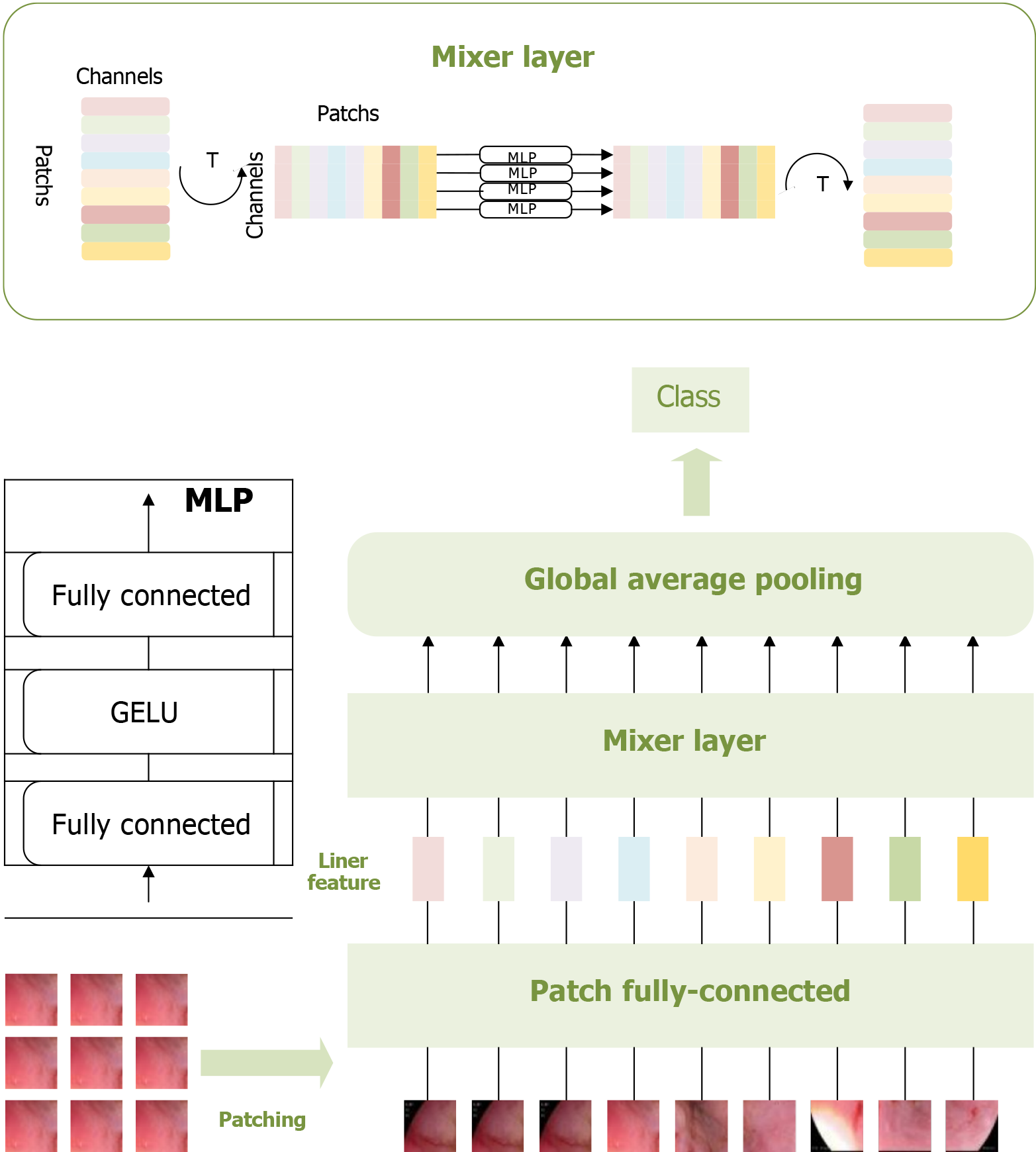
Figure 2 Multi-layer perceptron-based pathological recognition model for esophageal cancer.
This figure visualizes the patching, feature extraction, and classification process of esophageal adenocarcinoma images using the multi-layer perceptron-based model. These three operations constitute the core components of this model. Visualizing these processes provides valuable insights into the multi-layer perceptron model’s recognition performance, facilitating a comprehensive analysis of its effectiveness[23]. MLP: Multi-layer perceptron.
- Citation: Wei W, Zhang XL, Wang HZ, Wang LL, Wen JL, Han X, Liu Q. Application of deep learning models in the pathological classification and staging of esophageal cancer: A focus on Wave-Vision Transformer. World J Gastroenterol 2025; 31(19): 104897
- URL: https://www.wjgnet.com/1007-9327/full/v31/i19/104897.htm
- DOI: https://dx.doi.org/10.3748/wjg.v31.i19.104897


