Copyright
©The Author(s) 2024.
World J Gastroenterol. Jan 7, 2024; 30(1): 108-111
Published online Jan 7, 2024. doi: 10.3748/wjg.v30.i1.108
Published online Jan 7, 2024. doi: 10.3748/wjg.v30.i1.108
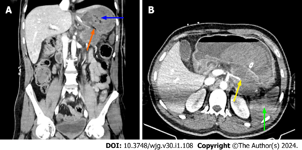
Figure 4 Computed tomography images of complicated necrotizing pancreatitis (original image).
A: Coronal image of the same patient (Figure 3) with walled off necrosis. Coronal computed tomography (CT) image showed that walled off necrosis was complicated by perforation into the stomach. A defect was seen on the wall of the necrotic collection (orange arrow). Stomach content was hyperdense (blue arrow) adjacent to the defect. When considered together with the gastrointestinal bleeding findings in the patient, this hyperdense appearance was thought to represent hemorrhage; B: Axial CT image showed that the splenic artery appeared to be occluded as it passes over the edge of the walled off necrosis (yellow arrow). It was also noteworthy that there was a near total loss of contrast enhancement in the spleen, consistent with infarction (green arrow).
- Citation: Ozturk MO, Aydin S. Complementary comments on diagnosis, severity and prognosis prediction of acute pancreatitis. World J Gastroenterol 2024; 30(1): 108-111
- URL: https://www.wjgnet.com/1007-9327/full/v30/i1/108.htm
- DOI: https://dx.doi.org/10.3748/wjg.v30.i1.108


