Copyright
©The Author(s) 2023.
World J Gastroenterol. Jan 21, 2023; 29(3): 561-578
Published online Jan 21, 2023. doi: 10.3748/wjg.v29.i3.561
Published online Jan 21, 2023. doi: 10.3748/wjg.v29.i3.561
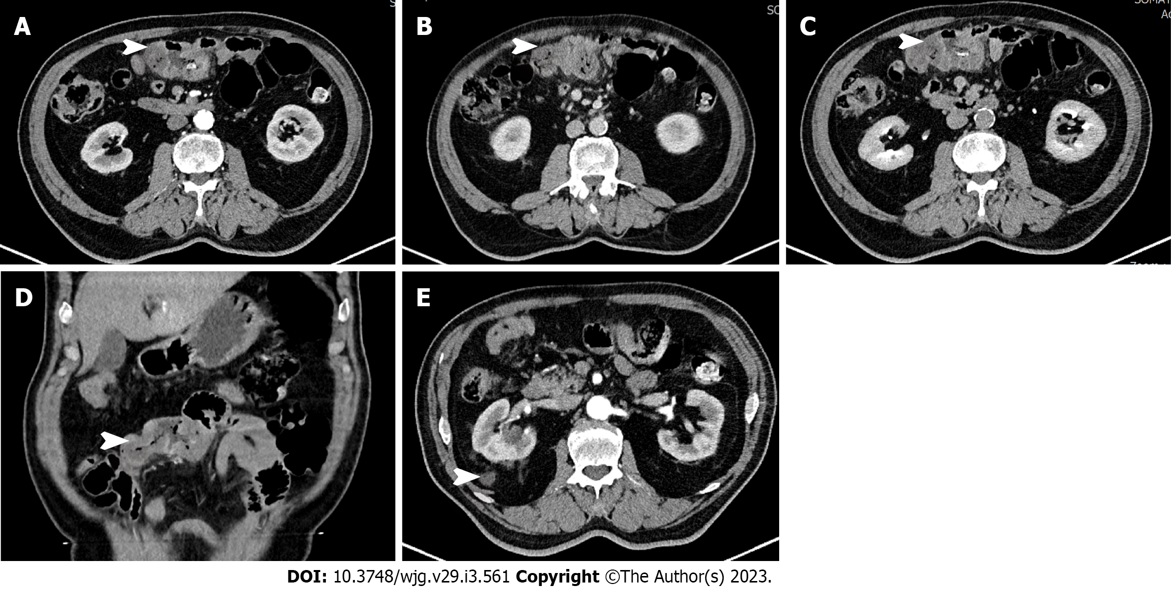
Figure 1 Computed tomography showed segmental thickening of the small intestine (white arrow), with lesion enhancement in the arterial phase.
A: Arterial phase; B: Venous phase; C: Balanced phase; D: Coronal plane; E: Adrenal masses.
- Citation: Ma XM, Yang BS, Yang Y, Wu GZ, Li YW, Yu X, Ma XL, Wang YP, Hou XD, Guo QH. Small intestinal angiosarcoma on clinical presentation, diagnosis, management and prognosis: A case report and review of the literature. World J Gastroenterol 2023; 29(3): 561-578
- URL: https://www.wjgnet.com/1007-9327/full/v29/i3/561.htm
- DOI: https://dx.doi.org/10.3748/wjg.v29.i3.561


