Case Report
Copyright ©The Author(s) 2022.
World J Gastroenterol. Nov 7, 2022; 28(41): 5993-6001
Published online Nov 7, 2022. doi: 10.3748/wjg.v28.i41.5993
Figure 2
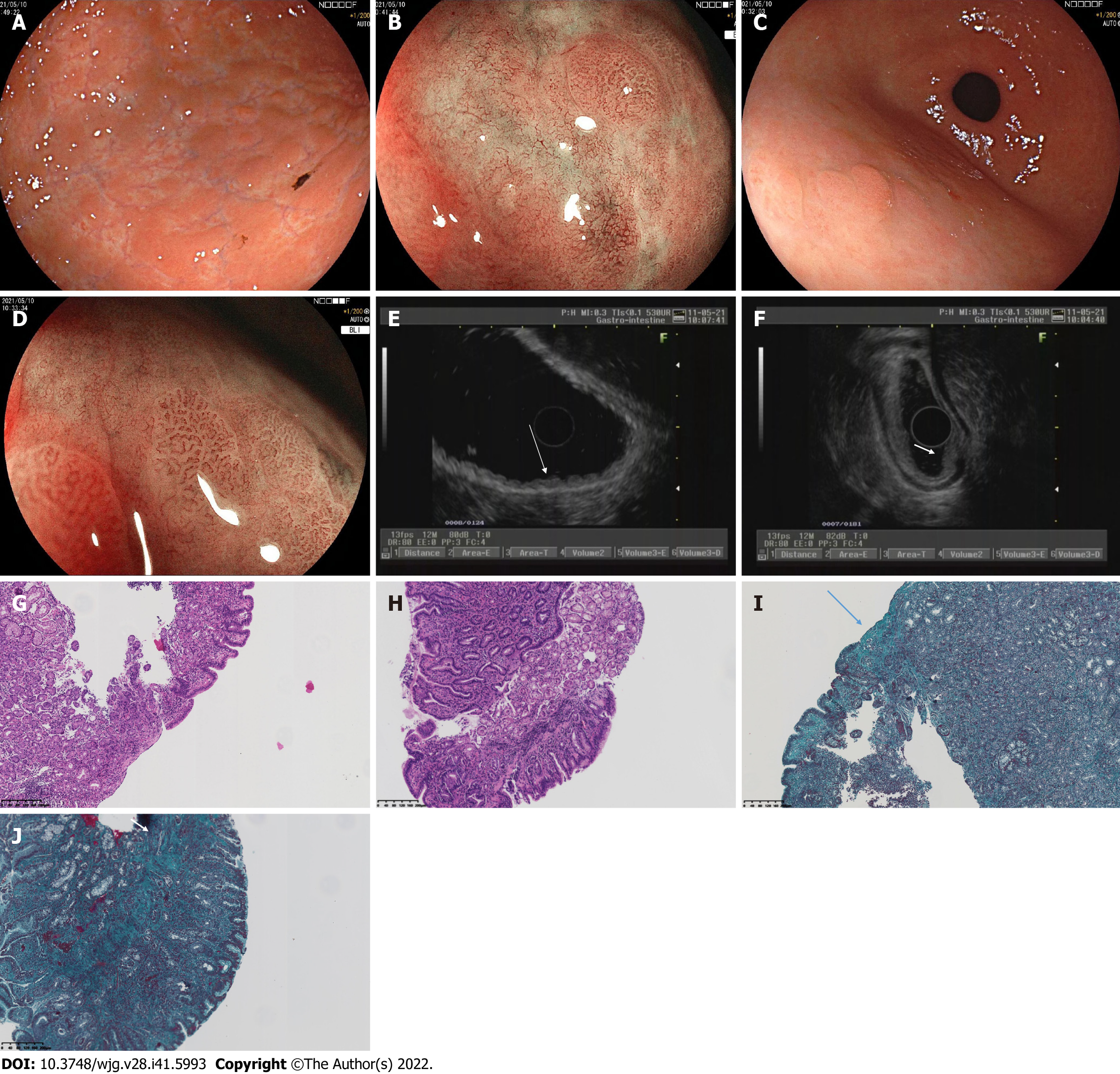
Figure 2 Findings of white light endoscopy, magnifying endoscopy, endoscopic ultrasound, and histopathology in 2021. A: Esophagogastroduodenoscopy (EGD) in 2021 showed that the mucosa of the angulus was smooth, while the mucosa of the gastric corpus and fundus appeared uneven, with elevation-depression changes; B: Magnifying endoscopy with blue laser imaging (ME-BLI) in 2021 showed regular arrangement of the marginal crypt epithelium (MCE) and widened microvessels in the elevated area of the corpus, and dendritic irregular microvascular pattern and unremarkable MCE in the depressed area of the corpus; C: EGD in 2021 showed nodular reddening-like changes in the anterior wall of the gastric antrum, which is similar to those in 2020; D: ME-BLI in 2021 showed similar findings in the gastric antrum to those in the corpus; E: Endoscopic ultrasound (EUS) in 2021 showed that the mucosal layer of the gastric corpus was thickened, exhibiting slightly hypoechoic and wavy changes; F: EUS in 2021 showed that the normal five-layer structures in the gastric antrum were replaced by inhomogeneous hypoechoic changes; G: Histopathological findings of the gastric corpus in 2021 (corresponding to Figure 2A and B) included moderate chronic inflammation with erosion, mild activity, a higher number of lymphocytes, and a few neutrophils and eosinophils (20-30/high-powered fields); H: Histopathological findings of the gastric antrum in 2021 (corresponding to Figure 2C and D) showed moderate chronic inflammation with erosion and mild activity; I: Masson staining of a gastric corpus specimen obtained in 2021 (corresponding to Figure 2G). The arrow indicates the collagen band, which had a thickness of about 40 μm; J: Masson staining of a gastric antrum specimen obtained in 2021 (corresponding to Figure 2H). The arrow also indicates the collagen band.